Monoclonal Antibodies
Anti-NUMA1 Rabbit Monoclonal Antibody for WB, IHC-P, ELISA - Q14980
Item Number : CM0011410
Price varies based on specs and customizations
- Application
- WB, IHC-P, ELISA
- Cross Reactivity
- Human, Mouse, Rat
- Protein Weight
- 238kDa

Shipping:
Contact us to get shipping details Enjoy On-time Dispatch Guarantee.
Why Choose Us
Easy ordering process, quality products, and dedicated support for your business success.
Core Product Specifications and Parameters
| Parameter | Value |
|---|---|
| Product Name | NUMA1 Rabbit mAb |
| Remarks/Alias | NUMA; NMP-22; NUMA1 |
| Species | Human |
| GeneID (Human) | 4926 |
| GeneID | 4926 |
| Immunogen | A synthetic peptide corresponding to a sequence within amino acids 2000-2100 of human NUMA11 (Q14980). |
| Source | Rabbit |
| Category | Monoclonal Antibodies |
| Application | WB, IHC-P, ELISA |
| Cross Reactivity | Human, Mouse, Rat |
| SWISS | Q14980 |
| Protein Weight | 238kDa |
| Shipping | Ice bag |
Biological Background: NUMA1 Function and Localization
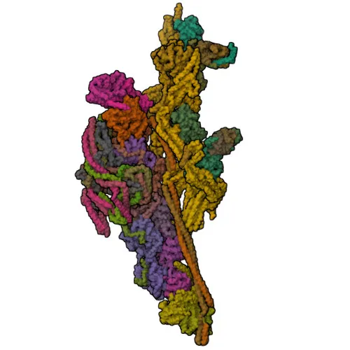
- NUMA1 (Nuclear Mitotic Apparatus Protein 1), also known as NMP-22 or NuMA, is a large coiled-coil protein essential for mitotic spindle organization and chromosome segregation.
- It tethers microtubule minus ends at spindle poles, which is critical for establishing and maintaining spindle pole integrity. Related references: PMID:11956313 PMID:12445386
- NUMA1 regulates spindle orientation and elongation during metaphase and anaphase via a dynein-dynactin-dependent mechanism, involving interactions with GPSM2 and G(i) alpha proteins at the cell cortex. Related references: PMID:22327364 PMID:23027904 PMID:23921553 PMID:24109598 PMID:23870127 PMID:24996901
- During anaphase, NUMA1 recruits dynein-dynactin to polar cortical regions through direct binding to phosphatidylinositol 4,5-bisphosphate (PI(4,5)P2), facilitating spindle elongation and chromosome segregation. Related references: PMID:24371089 PMID:22327364 PMID:23921553 PMID:24996901
- Beyond mitosis, NUMA1 is a major structural constituent of the nuclear matrix, occupying a large volume of the interphase nucleus and possibly serving a non-mitotic scaffolding role. Related references: PMID:10075938
- It is required for epidermal differentiation and hair follicle morphogenesis, indicating roles in tissue development.
- Subcellular localization dynamically shifts between nucleoplasm, spindle poles, centrosomes, cell cortex, and lateral cell membrane during the cell cycle.
- Post-translational modifications include acetylation, ADP-ribosylation, phosphorylation, and ubiquitination; it is also a lipid-binding lipoprotein and glycoprotein.
Experimental Guidance and Technical Tips
- The immunogen lies within the C-terminal region (aa 2000–2100) of human NUMA1; consider that the antibody may detect both full-length protein and possible C-terminal fragments.
- For Western blotting (WB), note the high molecular weight (~238 kDa); use low-percentage gels, extended transfer times, and confirm that protein ladders are properly resolved.
- In immunohistochemistry (IHC-P), expect nuclear staining in interphase cells and spindle-pole accumulation in mitotic cells; validate with appropriate cell-cycle markers or synchronized cultures.
- Cross-reactivity is indicated for human, mouse, and rat; verify reactivity in your target species and sample type before critical experiments.
- For ELISA applications, optimize coating concentrations and include relevant controls to ensure specificity.
CamelBio: Your One-Stop Sourcing Bridge
CamelBio provides a comprehensive range of IVD raw materials, customized antibody development, and technical support to streamline your diagnostic workflow. This anti-NUMA1 monoclonal antibody supports research in mitotic cell division and nuclear architecture, serving as a reliable tool for assay development. From validated antibody pairs and optimized monoclonals to bulk ancillary reagents, CamelBio helps bridge the gap between concept and clinic.
REQUEST A QUOTE
Our professional team will reply to you within one business day. Please feel free to contact us!