Polyclonal Antibodies
Anti-NSFL1C Rabbit Polyclonal Antibody (p47) for WB, IF/ICC, IP, ELISA - Q9UNZ2
Item Number : CM0025325
Price varies based on specs and customizations
- Application
- WB, IF/ICC, IP, ELISA
- Cross Reactivity
- Human, Mouse, Rat
- Protein Weight
- 41kDa

Shipping:
Contact us to get shipping details Enjoy On-time Dispatch Guarantee.
Why Choose Us
Easy ordering process, quality products, and dedicated support for your business success.
Core Product Specifications and Parameters
| Parameter | Value |
|---|---|
| Product Name | NSFL1C Rabbit pAb |
| Remarks/Alias | P47; UBX1; UBXD10; UBXN2C; dJ776F14.1 |
| Species | Human |
| Gene ID (Human) | 55968 |
| Gene ID | 55968 |
| Immunogen | Recombinant fusion protein containing a sequence corresponding to amino acids 1-185 of human NSFL1C (NP_057227.2) |
| Source | Rabbit |
| Category | Polyclonal Antibodies |
| Application | WB, IF/ICC, IP, ELISA |
| Cross Reactivity | Human, Mouse, Rat |
| SWISS | Q9UNZ2 |
| Protein Weight | 41kDa |
| Shipping | Ice bag |
Biological Background: NSFL1C Function and Localization
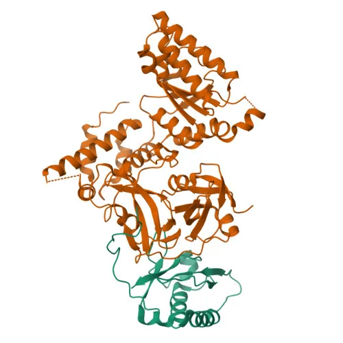
- NSFL1C, also known as p47, UBXN2C, or p97 cofactor p47, is a UBX domain-containing protein that serves as a cofactor for the AAA ATPase VCP/p97.
- It reduces the ATPase activity of VCP and is essential for mitotic Golgi stack fragmentation and subsequent VCP-mediated reassembly.
- Plays a role in VCP-mediated formation of transitional endoplasmic reticulum (tER).
- Inhibits the activity of cathepsin L (CTSL) in vitro. Related references: PMID:15498563
- Together with UBXN2B/p37, it regulates the centrosomal levels of kinase AURKA during mitotic progression by promoting AURKA removal from centrosomes in prophase, and also regulates spindle orientation. Related references: PMID:23649807
- Subcellular localization includes nucleus, Golgi apparatus (Golgi stack), chromosome, cytoplasm, cytoskeleton, and centrosome.
- The protein is subject to alternative splicing and phosphorylation, and features a lipid-binding UBX domain.
Experimental Guidance and Technical Tips
- The immunogen covers the N-terminal region (aa 1–185) of human NSFL1C. For western blotting, the predicted molecular weight is 41 kDa, but it is advisable to validate the observed band in your specific lysate system.
- For immunofluorescence and immunocytochemistry, permeabilization protocols may be critical to detect Golgi and centrosomal localizations; consider testing different fixation and permeabilization conditions.
- For immunoprecipitation, pre-clearing steps and appropriate control antibodies are recommended to minimize non-specific binding.
- Cross-reactivity with mouse and rat is reported; however, it is prudent to confirm sequence homology for your species of interest before use.
CamelBio: Your One-Stop Sourcing Bridge
CamelBio is your one-stop partner for IVD raw materials, offering validated antibody pairs, optimized monoclonal and polyclonal antibodies, and bulk ancillary reagents. For research into Golgi dynamics, mitosis, and VCP-related pathways, CamelBio supports sourcing of rare-target raw materials including NSFL1C/p47 antibodies. From concept to clinic, we bridge your diagnostic development needs with reliable supply solutions.
Product Datasheet
Anti-NSFL1C Rabbit Polyclonal Antibody (p47) for WB, IF/ICC, IP, ELISA - Q9UNZ2
REQUEST A QUOTE
Our professional team will reply to you within one business day. Please feel free to contact us!