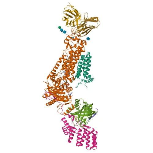
Monoclonal Antibodies
Anti-NOXA2/p67phox Rabbit Monoclonal Antibody for WB, IHC-P, ELISA - P19878
Item Number : CM0012020
Price varies based on specs and customizations
- Application
- WB, IHC-P, ELISA
- Cross Reactivity
- Mouse, Rat
- Protein Weight
- 60kDa

Shipping:
Contact us to get shipping details Enjoy On-time Dispatch Guarantee.
Why Choose Us
Easy ordering process, quality products, and dedicated support for your business success.
Core Product Specifications and Parameters
| Parameter | Value |
|---|---|
| Product Name | NOXA2/p67phox Rabbit mAb |
| Remarks/Alias | NCF-2, NOXA2, P67PHOX, P67-PHOX, NOXA2/p67phox |
| Species | Human |
| GeneID (Human) | 4688 |
| GeneID | 4688 |
| Immunogen | Recombinant fusion protein containing a sequence corresponding to amino acids 200-300 of human NOXA2/p67phox (P19878). |
| Source | Rabbit |
| Category | Monoclonal Antibodies |
| Application | WB, IHC-P, ELISA |
| Cross Reactivity | Mouse, Rat |
| SWISS | P19878 |
| Protein Weight | 60kDa |
| Shipping | Ice bag |
Biological Background: NOXA2/p67phox Function and Localization
- NCF2/p67-phox is a key cytosolic subunit of the phagocyte NADPH oxidase complex, which transfers electrons from NADPH to molecular oxygen to generate superoxide, essential for microbial killing. Related references: PMID:12207919 PMID:38355798
- Activation of the oxidase involves phosphorylation-dependent assembly of NCF1/p47-phox with the membrane-bound CYBA/p22-phox, recruiting the cytosolic complex to the nascent phagosome.
- The protein localizes predominantly to the cytoplasm and translocates to membranes upon stimulation.
- Structural domains include an N-terminal TPR repeat and a C-terminal SH3 domain, which mediate protein–protein interactions critical for complex formation.
- Alternative splicing produces multiple isoforms, potentially diversifying regulatory functions.
- Mutations in NCF2 are linked to chronic granulomatous disease (CGD), a primary immunodeficiency characterized by recurrent infections due to defective superoxide production.
Experimental Guidance and Technical Tips
- The immunogen (amino acids 200–300) lies within a central region of p67-phox that is conserved between human, mouse, and rat, consistent with the reported cross-reactivity.
- For Western blot, the predicted molecular weight is ~60 kDa; consider using appropriate positive controls (e.g., neutrophil or macrophage lysates) and validate the signal with appropriate inhibitors or siRNA.
- In IHC-P, antigen retrieval optimization may be required; verify staining specificity on tissues known to express high levels of phagocyte oxidase components, such as spleen or inflamed tissues.
- Due to the monoclonal nature, epitope specificity is consistent, but always confirm reactivity with your target cells under physiological and stimulated conditions.
CamelBio: Your One-Stop Sourcing Bridge
CamelBio is a dedicated partner for IVD raw-material procurement, connecting you with reliable antibodies, antigen reagents, and ancillary products across diverse research areas. For phagocyte oxidase studies, we offer validated antibody pairs, optimized monoclonal/polyclonal antibodies, and bulk ancillary reagents to support assay development. Our global sourcing network can also assist with hard-to-find targets, ensuring a streamlined supply chain from concept to clinic.
Product Datasheet
Anti-NOXA2/p67phox Rabbit Monoclonal Antibody for WB, IHC-P, ELISA - P19878
REQUEST A QUOTE
Our professional team will reply to you within one business day. Please feel free to contact us!