Monoclonal Antibodies
Anti-Notch1 Monoclonal Antibody for WB, IP, ELISA - P46531
Item Number : CM0003362
Price varies based on specs and customizations
- Application
- WB, IP, ELISA
- Cross Reactivity
- Human, Mouse
- Protein Weight
- 273kDa

Shipping:
Contact us to get shipping details Enjoy On-time Dispatch Guarantee.
Why Choose Us
Easy ordering process, quality products, and dedicated support for your business success.
Core Product Specifications and Parameters
| Parameter | Value |
|---|---|
| Product Name | Notch1 Rabbit mAb |
| Remarks/Alias | hN1; AOS5; TAN1; AOVD1; Notch1 |
| Species | Human |
| GeneID (Human) | 4851 |
| GeneID | 4851 |
| Immunogen | Synthetic Peptide|A synthetic peptide corresponding to a sequence within amino acids 2456-2555 of human Notch1 (P46531). |
| Source | Rabbit |
| Category | Monoclonal Antibodies |
| Application | WB, IP, ELISA |
| Cross Reactivity | Human, Mouse |
| SWISS | P46531 |
| Protein Weight | 273kDa |
| Shipping | Ice bag |
Biological Background: Notch1 Function and Localization
- Neurogenic locus notch homolog protein 1 (Notch1) is a transmembrane receptor that mediates cell-cell communication to regulate cell-fate decisions, differentiation, proliferation, and apoptosis.
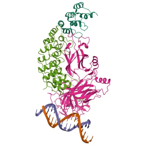
- Functions as a receptor for membrane-bound ligands Jagged-1, Jagged-2, and Delta-1; upon ligand binding, the Notch intracellular domain (NICD) is released and translocates to the nucleus, where it forms a transcriptional activator complex with RBPJ to drive target gene expression.
- Involved in angiogenesis by negatively regulating endothelial cell proliferation, migration, and angiogenic sprouting.
- Essential for T-cell maturation in the thymus, contributing to the development of both CD4+ and CD8+ cells.
- Subcellular localization includes the cell membrane, late endosome membrane, and nucleus, reflecting its proteolytic activation and signaling dynamics.
- In fetal tissues, most abundant in spleen, brain stem, and lung; in adults, predominantly found in lymphoid tissues.
- Post-translational modifications include phosphorylation, glycosylation, hydroxylation, and ubiquitination, as indicated by keywords.
- Plays a role in left/right symmetry determination and cilia function at the left-right organizer.
Experimental Guidance and Technical Tips
- The immunogen lies within the intracellular domain (aa 2456-2555), so the antibody should recognize both full-length Notch1 and the released NICD. Verify detection of expected bands in Western blot.
- For WB, use a low-percentage polyacrylamide gel (e.g., 6-8%) to resolve the high molecular weight protein (~273 kDa). Prolonged transfer may be required.
- Cross-reactivity with mouse Notch1 is reported; validate detection in mouse tissue or cell lysates if needed.
- For IP, consider mild lysis conditions to maintain protein-protein interactions if studying complexes.
- As a rabbit monoclonal antibody, expect high specificity and lot-to-lot consistency; always titrate for optimal dilution in your assays.
CamelBio: Your One-Stop Sourcing Bridge
CamelBio offers diagnostic manufacturers, laboratories, and research institutes a one-stop sourcing platform for IVD raw materials, including validated antibody pairs, optimized monoclonal and polyclonal antibodies, and bulk ancillary reagents. For researchers investigating Notch signaling, cell fate determination, and developmental disorders, our supply chain supports access to high-quality antibodies like this anti-Notch1 clone. We also specialize in sourcing raw materials for rare targets to accelerate your assay development from concept to clinic.
REQUEST A QUOTE
Our professional team will reply to you within one business day. Please feel free to contact us!