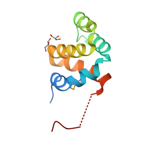
Polyclonal Antibodies
Anti-NOL3 Polyclonal Antibody for WB, IF/ICC, ELISA - O60936
Item Number : CM0015305
Price varies based on specs and customizations
- Application
- WB, IF/ICC, ELISA
- Cross Reactivity
- Human, Mouse
- Protein Weight
- 23kDa

Shipping:
Contact us to get shipping details Enjoy On-time Dispatch Guarantee.
Why Choose Us
Easy ordering process, quality products, and dedicated support for your business success.
Core Product Specifications and Parameters
| Parameter | Value |
|---|---|
| Product Name | NOL3 Rabbit pAb |
| Remarks/Alias | ARC; FCM; MYP; NOP; NOP30; MYOCL1; NOL3 |
| Species | Human |
| Gene ID (Human) | 8996 |
| Gene ID | 8996 |
| Immunogen | Recombinant fusion protein containing a sequence corresponding to amino acids 1-208 of human NOL3 (NP_003937.1) |
| Source | Rabbit |
| Category | Polyclonal Antibodies |
| Application | WB, IF/ICC, ELISA |
| Cross Reactivity | Human, Mouse |
| SWISS | O60936 |
| Protein Weight | 23kDa |
| Shipping | Ice bag |
Biological Background: NOL3 Function and Localization
- NOL3, also known as ARC, FCM, MYP, or NOP30, is a nucleolar protein that functions as an apoptosis repressor, blocking multiple cell death pathways.
- It inhibits extrinsic apoptosis by interacting with FAS and FADD to block DISC assembly, and by sequestering CASP8 in a phosphorylation-dependent manner at mitochondria.
- In the intrinsic pathway, NOL3 binds to BAX, preventing its activation, mitochondrial dysfunction, and release of pro-apoptotic factors. Related references: PMID:15004034
- NOL3 acts as a cytosolic calcium buffer, disrupting the CASP8 interaction and preserving calcium homeostasis to inhibit calcium-mediated cell death. Related references: PMID:15509781
- It negatively regulates apoptosis induced by oxidative stress and hypoxia by blocking CASP2 activation, BAX translocation, and cytochrome c release.
- NOL3 also suppresses TNF-induced necrosis by interacting with TNFRSF1A and preventing RIPK1 recruitment to complex I.
- Subcellularly, NOL3 localizes to the nucleus, nucleolus, cytoplasm, mitochondria, sarcoplasmic reticulum, and membrane, reflecting its multifunctional roles.
- Highly expressed in heart and skeletal muscle, with lower levels in placenta, liver, kidney, and pancreas, suggesting tissue-specific apoptosis regulation.
Experimental Guidance and Technical Tips
- For WB, the expected band is at ~23 kDa; consider using appropriate positive and negative controls to validate specificity.
- In IF/ICC, NOL3 exhibits a complex localization pattern; ensure fixation and permeabilization conditions are optimized for your cell type and compartment of interest.
- For ELISA, a recombinant NOL3 protein standard is recommended for quantitative assessments; cross-reactivity with mouse NOL3 should be verified in your assay system.
- As a polyclonal antibody, it may recognize multiple epitopes; lot-to-lot consistency should be monitored in critical applications.
CamelBio: Your One-Stop Sourcing Bridge
CamelBio provides diagnostic manufacturers and research teams with integrated access to high-quality IVD raw materials, including validated antibodies like this anti-NOL3 polyclonal antibody for apoptosis research. Whether you need bulk quantities of polyclonal antibodies, matched antibody pairs, or ancillary reagents, our streamlined sourcing platform supports your workflow from early-stage development to clinical application.
REQUEST A QUOTE
Our professional team will reply to you within one business day. Please feel free to contact us!