Monoclonal Antibodies
Anti-NMDAR1 Monoclonal Antibody for WB, IF-P, IHC-P, ELISA - Q05586
Item Number : CM0008636
Price varies based on specs and customizations
- Application
- WB, IF-P, IHC-P, ELISA
- Cross Reactivity
- Human, Mouse, Rat
- Protein Weight
- 105kDa

Shipping:
Contact us to get shipping details Enjoy On-time Dispatch Guarantee.
Why Choose Us
Easy ordering process, quality products, and dedicated support for your business success.
Core Product Specifications and Parameters
| Parameter | Value |
|---|---|
| Product Name | NMDAR1 Rabbit mAb |
| Remarks/Alias | NR1; MRD8; GluN1; NMDA1; DEE101; NDHMSD; NDHMSR; NMD-R1; NMDAR1 |
| Species | Human |
| Gene ID (Human) | 2902 |
| Immunogen | Synthetic peptide |
| Source | Rabbit |
| Category | Monoclonal Antibodies |
| Application | WB, IF-P, IHC-P, ELISA |
| Cross Reactivity | Human, Mouse, Rat |
| SWISS | Q05586 |
| Protein Weight | 105kDa |
| Shipping | Ice bag |
Biological Background: NMDAR1 Function and Localization
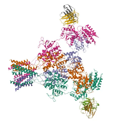
- NMDAR1 (GRIN1) encodes the glutamate receptor ionotropic NMDA 1 subunit, also known as GluN1, NR1, or NMD-R1. The human protein (UniProt: Q05586) is a 105 kDa glycoprotein with predicted alternative splicing that generates multiple isoforms.
- Forms the obligate subunit of N-methyl-D-aspartate (NMDA) receptors, functioning as heterotetrameric, ligand-gated cation channels with high calcium permeability and voltage-dependent block by Mg²⁺. Activation requires simultaneous binding of glutamate to GluN2 subunits and glycine/D-serine to GluN1, plus membrane depolarization. Related references: PMID:21376300, PMID:26875626, PMID:26919761
- Critical for synaptic plasticity, learning, and memory formation through long-term potentiation (LTP). Related references: PMID:26875626
- Subunit composition determines channel kinetics: GluN2 and GluN3 subunits modulate activation, deactivation, desensitization, pH sensitivity, Ca²⁺ permeability, and allosteric modulator binding. Related references: PMID:26875626, PMID:26919761, PMID:36309015
- Localized predominantly to the postsynaptic membrane, particularly at postsynaptic density, where it mediates simultaneous potassium efflux and calcium/sodium influx.
- Subject to extensive post-translational modifications including phosphorylation, N-glycosylation, and ubiquitination, which regulate receptor trafficking and function.
- Associated with neurological disorders: gene mutations cause developmental and epileptic encephalopathy (DEE101), intellectual disability, and other neurodevelopmental phenotypes.
- Contains multiple functional domains: extracellular ligand-binding domain, transmembrane helices, and intracellular C-terminal domain involved in scaffolding and signaling.
Experimental Guidance and Technical Tips
- This antibody was generated against a synthetic peptide spanning amino acids 601-700 of human NMDAR1 (UniProt Q05586). This region lies within the intracellular C-terminal domain. Consider using denaturing gel electrophoresis for WB, as the epitope may be linear.
- For immunofluorescence or immunohistochemistry, verify staining specificity by using appropriate controls (e.g., peptide blocking or knockout tissue).
- In ELISA applications, the antibody may be used as a capture or detection reagent; optimal dilutions should be empirically determined.
CamelBio: Your One-Stop Sourcing Bridge
CamelBio serves as a one-stop sourcing platform for IVD raw materials, offering validated antibody reagents, bulk ancillary materials, and custom development services. For NMDA receptor research and diagnostic applications targeting neurological disorders, CamelBio can supply monoclonal antibodies, supporting your assay development and commercialization needs.
REQUEST A QUOTE
Our professional team will reply to you within one business day. Please feel free to contact us!