Monoclonal Antibodies
Anti-NF-κB2 Monoclonal Antibody for WB, IHC-P, ELISA - Q00653
Item Number : CM0003763
Price varies based on specs and customizations
- Application
- WB, IHC-P, ELISA
- Cross Reactivity
- Human, Mouse, Rat
- Protein Weight
- 97kDa

Shipping:
Contact us to get shipping details Enjoy On-time Dispatch Guarantee.
Why Choose Us
Easy ordering process, quality products, and dedicated support for your business success.
Core Product Specifications and Parameters
| Parameter | Value |
|---|---|
| Product Name | NF-κB2 Rabbit mAb |
| Remarks/Alias | p52; p100; H2TF1; LYT10; CVID10; LYT-10; NF-kB2; p49/p100; NF-κB2 |
| Species | Human |
| GeneID (Human) | 4791 |
| GeneID | 4791 |
| Immunogen | Recombinant fusion protein containing a sequence corresponding to amino acids 745-877 of human NF-κB2 (Q00653) |
| Source | Rabbit |
| Category | Monoclonal Antibodies |
| Application | WB, IHC-P, ELISA |
| Cross Reactivity | Human, Mouse, Rat |
| SWISS | Q00653 |
| Protein Weight | 97kDa |
| Shipping | Ice bag |
Biological Background: NF-κB2 Function and Localization
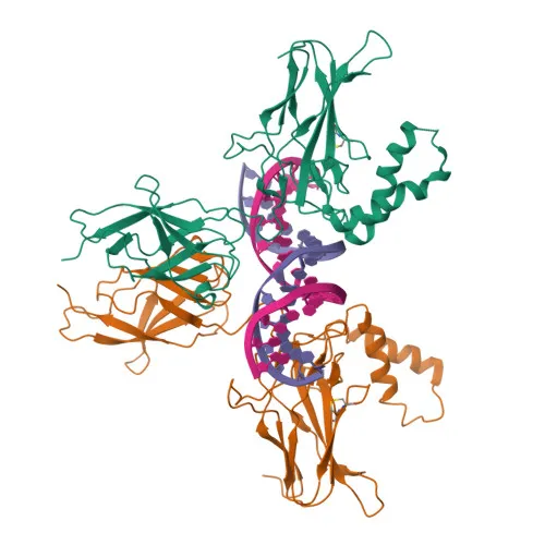
- NF-κB2 encodes the p100 subunit, which undergoes proteolytic processing to generate the active DNA-binding p52 subunit; isoform p49 is also produced by alternative splicing.
- p100/p52 belongs to the NF-kappa-B transcription factor family, forming homo- or heterodimers with Rel-like proteins (RELA, RELB, REL, NFKB1) to regulate gene expression.
- In the non-canonical NF-kappa-B pathway, p100 associated with RelB is phosphorylated by IKKα and processed to p52, leading to nuclear translocation of the RelB-p52 transcriptional activator complex.
- p52 homodimers act as transcriptional repressors, while RelB-p52 heterodimers activate transcription, binding to kappa-B sites (5'-GGRNNYYCC-3') in promoters of immune and inflammatory genes.
- NF-κB2 is involved in a wide range of biological processes including inflammation, immunity, cell growth, differentiation, and apoptosis.
- In concert with RELB, NF-κB2 regulates the circadian clock by repressing the transcriptional activator activity of the CLOCK-BMAL1 heterodimer.
- Subcellular localization is both cytoplasmic (as inactive p100/I-kappa-B complexes) and nuclear (as active p52-containing dimers).
- Ank repeats, isopeptide bond, and ubl conjugation (SUMOylation) are post-translational modifications, and the protein is also phosphorylated.
Experimental Guidance and Technical Tips
- The immunogen corresponds to amino acids 745-877 within the C-terminal ankyrin repeat domain of p100, which is absent in the processed p52 subunit; therefore, the antibody may preferentially recognize full-length p100 (~97 kDa). Consider validating reactivity against both p100 and p52 in your model system if required.
- For western blotting, use standard SDS-PAGE and transfer conditions; a band near 97 kDa is expected for p100. Lower bands may represent processing intermediates or degradation products.
- In immunohistochemistry (IHC-P), both nuclear and cytoplasmic staining may be observed depending on the activation status of NF-κB2 in the tissue. Include appropriate controls for signal specificity.
- For ELISA, the antibody may be used with recombinant p100 or cell lysates; ensure the coating antigen contains the C-terminal epitope region.
- Cross-reactivity with mouse and rat suggests conservation of the immunogenic sequence among these species, but sequence alignment is recommended to confirm epitope identity.
CamelBio: Your One-Stop Sourcing Bridge
CamelBio supports diagnostic manufacturers and research laboratories with comprehensive IVD raw-material sourcing, technical services, and consulting—from concept to clinic. For NF-κB2 and other NF-kappa-B pathway targets, suppliers on CamelBio can provide validated antibody pairs, optimized monoclonal and polyclonal antibodies, bulk ancillary reagents, and assistance with rare target raw-material procurement.
REQUEST A QUOTE
Our professional team will reply to you within one business day. Please feel free to contact us!