Monoclonal Antibodies
Anti-NF-κB2 Monoclonal Antibody for WB, ELISA - Q00653
Item Number : CM0004541
Price varies based on specs and customizations
- Application
- WB, ELISA
- Cross Reactivity
- Human, Mouse, Rat
- Protein Weight
- 97kDa

Shipping:
Contact us to get shipping details Enjoy On-time Dispatch Guarantee.
Why Choose Us
Easy ordering process, quality products, and dedicated support for your business success.
Core Product Specifications and Parameters
| Parameter | Value |
|---|---|
| Product Name | NF-κB2 Rabbit mAb |
| Remarks/Alias | p52; p100; H2TF1; LYT10; CVID10; LYT-10; NF-kB2; p49/p100; NF-κB2 |
| Species | Human |
| GeneID (Human) | 4791 |
| GeneID | 4791 |
| Immunogen | Synthetic Peptide - A synthetic peptide corresponding to a sequence within amino acids 800-900 of human NF-κB2 (NP_001070962.1) |
| Source | Rabbit |
| Category | Monoclonal Antibodies |
| Application | WB, ELISA |
| Cross Reactivity | Human, Mouse, Rat |
| SWISS | Q00653 |
| Protein Weight | 97kDa |
| Shipping | Ice bag |
Biological Background: NF-κB2 Function and Localization
- NF-κB2 encodes the p100 precursor and its proteolytically processed p52 subunit, both of which function as components of the NF-κB transcription factor complex.
- The p52-RELB heterodimer acts as a transcriptional activator, while the p52-p52 homodimer functions as a repressor, illustrating the dimer-specific regulation of NF-κB target genes.
- NF-κB is a pleiotropic transcription factor that responds to diverse stimuli and controls genes involved in inflammation, immunity, cell growth, differentiation, and apoptosis.
- Full-length p100 retains associated NF-κB proteins in the cytoplasm via its ankyrin repeat domain; upon non-canonical pathway activation, proteasome-mediated processing generates p52, which translocates to the nucleus.
- An alternative isoform (p49) can stimulate the HIV enhancer in synergy with p65 (RELA).
- NF-κB2, together with RELB, regulates the circadian clock by repressing the transcriptional activity of the CLOCK-BMAL1 heterodimer.
- Post-translational modifications such as phosphorylation, ubiquitination, and processing are critical for NF-κB2 activity, as reflected in keywords like "Phosphoprotein" and "Isopeptide bond".
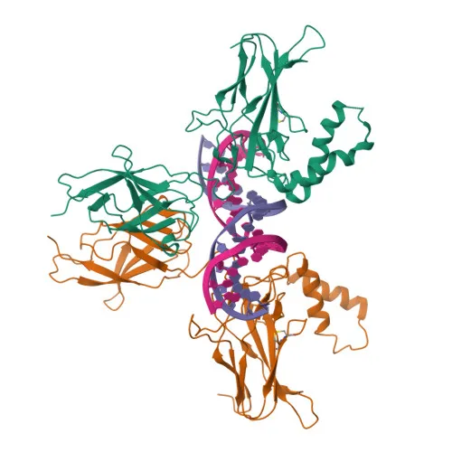
- The protein contains ankyrin repeats and a DNA-binding Rel homology domain, enabling its dual roles in cytoplasmic retention and transcriptional regulation.
Experimental Guidance and Technical Tips
- The immunogen maps to a C-terminal region (aa 800–900) within the p100-specific ankyrin repeat domain; this antibody is expected to detect full-length p100 (~97 kDa) but not the processed p52 subunit.
- For Western blot detection, consider analyzing whole-cell lysates from populations stimulated with NF-κB pathway activators (e.g., TNF-α, LIGHT) to enhance p100 signals.
- Reported cross-reactivity with mouse and rat samples; validate the antibody in the relevant species and tissue context by including appropriate positive and negative controls.
- In ELISA applications, optimize coating antigen and antibody titration under your assay conditions to achieve desired sensitivity and specificity.
CamelBio: Your One-Stop Sourcing Bridge
CamelBio provides diagnostic manufacturers, labs, and research institutes with one-stop access to IVD raw materials, technical services, and consulting—covering every stage from concept to clinic. As a sourcing partner for NF-κB pathway research, we offer validated monoclonal antibodies, optimized antibody pairs, and bulk ancillary reagents to support immunoassay development and rare target procurement. Our portfolio helps streamline your workflow in inflammation, oncology, and immune signaling studies, combining flexible supply with expert guidance.
REQUEST A QUOTE
Our professional team will reply to you within one business day. Please feel free to contact us!