Monoclonal Antibodies
Anti-NDUFA1 Rabbit Monoclonal Antibody for WB, IHC-P, ELISA - O15239
Item Number : CM0002622
Price varies based on specs and customizations
- Application
- WB, IHC-P, ELISA
- Cross Reactivity
- Human
- Protein Weight
- 8kDa

Shipping:
Contact us to get shipping details Enjoy On-time Dispatch Guarantee.
Why Choose Us
Easy ordering process, quality products, and dedicated support for your business success.
Core Product Specifications and Parameters
| Parameter | Value |
|---|---|
| Product Name | NDUFA1 Rabbit mAb |
| Remarks/Alias | MWFE; ZNF183; CI-MWFE; MC1DN12; NDUFA1 |
| Species | Human |
| GeneID (human) | 4694 |
| GeneID | 4694 |
| Immunogen | Synthetic peptide|A synthetic peptide corresponding to a sequence within amino acids 1-70 of human NDUFA1 (O15239). |
| Source | Rabbit |
| Category | Monoclonal Antibodies |
| Application | WB, IHC-P, ELISA |
| Cross Reactivity | Human |
| SWISS | O15239 |
| Protein Weight | 8kDa |
| Shipping | Ice bag |
Biological Background: NDUFA1 Function and Localization
- NDUFA1 encodes the NADH dehydrogenase [ubiquinone] 1 alpha subcomplex subunit 1, also known as Complex I-MWFE (CI-MWFE) and NADH-ubiquinone oxidoreductase MWFE subunit.
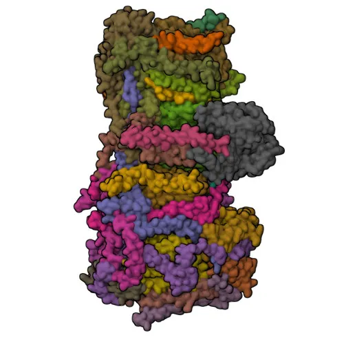
- It is an accessory subunit of mitochondrial respiratory chain Complex I, believed not to be involved in catalysis, but participates in the electron transfer from NADH to the respiratory chain, with ubiquinone as the immediate electron acceptor.
- Complex I is the first enzyme of the respiratory chain, coupling electron transfer from NADH to ubiquinone with proton translocation across the inner mitochondrial membrane.
- NDUFA1 is an integral membrane protein localized to the mitochondrial inner membrane, consistent with its role in the respiratory chain.
- Expression is primarily in heart and skeletal muscle, tissues with high energy demands.
- Disease variants in NDUFA1 are associated with primary mitochondrial diseases, particularly mitochondrial complex I deficiency.
- The protein has a calculated molecular weight of approximately 8 kDa and is predicted to contain transmembrane helices, as highlighted by keywords such as “Transmembrane” and “Transmembrane helix.”
Experimental Guidance and Technical Tips
- The immunogen was designed as a synthetic peptide spanning amino acids 1–70 of human NDUFA1. When using this antibody, consider the target’s mitochondrial localization and small size (8 kDa) for sample preparation and detection.
- For Western blotting, a high-percentage gel (e.g., 15% or gradient) and appropriate low-molecular-weight markers are advisable to resolve the ~8 kDa band.
- IHC-P signals should be validated in positive control tissues such as human heart or skeletal muscle, where NDUFA1 expression is abundant.
- In ELISA, the immunizing peptide may serve as a positive control for assay optimization, but each experimental system should be validated independently.
CamelBio: Your One-Stop Sourcing Bridge
CamelBio supports diagnostic manufacturers and research laboratories with a comprehensive portfolio of IVD raw materials. This anti-NDUFA1 monoclonal antibody is tailored for mitochondrial research and respiratory chain studies, offering reliable performance in WB, IHC-P, and ELISA. Through our one-stop sourcing model, we deliver validated antibody pairs, optimized monoclonal and polyclonal antibodies, bulk ancillary reagents, and even rare target raw materials to accelerate your project from concept to clinic.
REQUEST A QUOTE
Our professional team will reply to you within one business day. Please feel free to contact us!