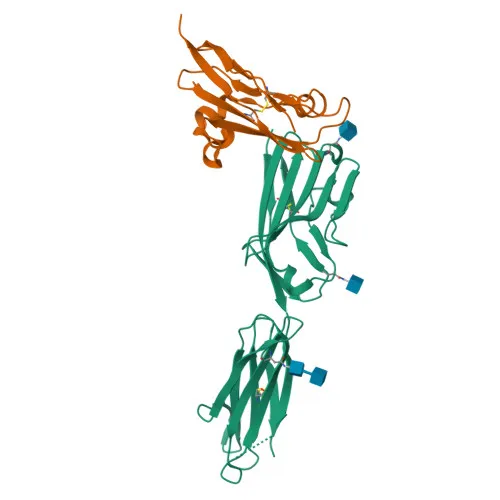
Monoclonal Antibodies
Anti-NCR3 Rabbit Monoclonal Antibody for WB, IF/ICC, FC, ELISA - O14931
Item Number : CM0007195
Price varies based on specs and customizations
- Application
- WB, IF/ICC, FC (intra), ELISA
- Cross Reactivity
- Human
- Protein Weight
- 16-21 kDa

Shipping:
Contact us to get shipping details Enjoy On-time Dispatch Guarantee.
Why Choose Us
Easy ordering process, quality products, and dedicated support for your business success.
Core Product Specifications and Parameters
| Parameter | Value |
|---|---|
| Product Name | NCR3 Rabbit mAb |
| Remarks/Alias | NCR3; 1C7; CD337; LY117; MALS; NKp30; natural cytotoxicity triggering receptor 3 |
| Species | Human |
| GeneID (Human) | 259197 |
| GeneID | 259197 |
| Immunogen | Recombinant protein |
| Source | Rabbit |
| Category | Monoclonal Antibodies |
| Application | WB, IF/ICC, FC (intra), ELISA |
| Cross Reactivity | Human |
| SWISS | O14931 |
| Protein Weight | 16kDa/17kDa/18kDa/19kDa/20kDa/21kDa |
| Shipping | Ice bag |
Biological Background: NCR3 Function and Localization
- NCR3, also designated NKp30 or CD337, is a member of the natural cytotoxicity receptor family and belongs to the immunoglobulin superfamily.
- It functions as an activating receptor selectively expressed on natural killer (NK) cells, where it recognizes stress-induced ligands BAG6 and NCR3LG1 on target cells.
- Ligand binding triggers NK cell cytotoxicity, playing a pivotal role in the immune surveillance against tumor cells.
- Beyond direct cytotoxicity, engagement of NCR3 by BAG6 promotes myeloid dendritic cell maturation through the release of TNFα and IFNγ by NK cells.
- The protein is localized to the cell membrane and features an extracellular immunoglobulin domain, a single transmembrane helix, and a short cytoplasmic tail.
- Post-translational modifications include disulfide bond formation and glycosylation, which may influence its molecular weight and functional properties.
- Alternative splicing generates multiple isoforms, contributing to the observed molecular weight range (16–21 kDa) in experimental settings.
- Tissue expression is largely restricted to resting and activated NK cells, with weak expression detected in the spleen.
Experimental Guidance and Technical Tips
- The immunogen spans the extracellular domain (amino acids 19–135), suggesting the antibody may recognize an epitope accessible in live cell applications; however, it is validated for intracellular flow cytometry—optimization for surface staining may be considered.
- Due to alternative splicing and glycosylation, expect multiple bands or bands between 16 and 21 kDa in WB; validate specificity using positive controls (e.g., NK cell lysates) and include appropriate blocking steps.
- For immunofluorescence and flow cytometry, inclusion of permeabilization and fixation steps is recommended in line with the validated intracellular staining protocol.
CamelBio: Your One-Stop Sourcing Bridge
For researchers and diagnostic developers working on NK cell biology and innate immunity, CamelBio streamlines access to critical raw materials such as Anti-NCR3 antibodies. Our portfolio includes validated antibody pairs, optimized monoclonal/polyclonal antibodies, and a wide range of ancillary reagents, supporting your IVD assay development from early-stage research to commercial production. Leverage our expertise to source even rare-target biomaterials efficiently under one roof.
Product Datasheet
Anti-NCR3 Rabbit Monoclonal Antibody for WB, IF/ICC, FC, ELISA - O14931
REQUEST A QUOTE
Our professional team will reply to you within one business day. Please feel free to contact us!