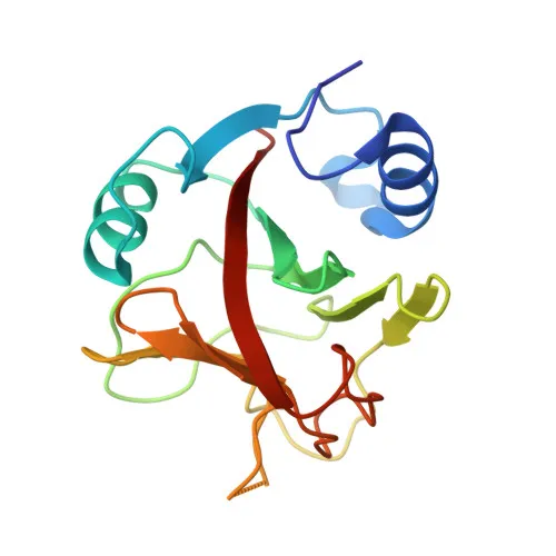
Polyclonal Antibodies
Anti-NCOA7 Polyclonal Antibody for WB, ELISA - Q8NI08
Item Number : CM0013520
Price varies based on specs and customizations
- Application
- WB, ELISA
- Cross Reactivity
- Human, Mouse, Rat
- Protein Weight
- 106kDa

Shipping:
Contact us to get shipping details Enjoy On-time Dispatch Guarantee.
Why Choose Us
Easy ordering process, quality products, and dedicated support for your business success.
Core Product Specifications and Parameters
| Parameter | Value |
|---|---|
| Product Name | NCOA7 Rabbit pAb |
| Remarks/Alias | ESNA1; TLDC4; ERAP140; NCOA7-AS; Nbla00052; Nbla10993; dJ187J11.3; NCOA7 |
| Species | Human |
| GeneID (Human) | 135112 |
| GeneID | 135112 |
| Immunogen | Recombinant protein: Recombinant fusion protein containing a sequence corresponding to amino acids 1-90 of human NCOA7 (NP_861447.3). |
| Source | Rabbit |
| Category | Polyclonal Antibodies |
| Application | WB, ELISA |
| Cross Reactivity | Human, Mouse, Rat |
| SWISS | Q8NI08 |
| Protein Weight | 106kDa |
| Shipping | Ice bag |
Biological Background: NCOA7 Function and Localization
- Nuclear receptor coactivator 7 (NCOA7) is a transcriptional coactivator that enhances the activities of several nuclear receptors, including ESR1, THRB, PPARG, and RARA. Related references: PMID:11971969
- It modulates vacuolar-type H+-ATPase (V-ATPase) activity by inducing disassembly of the V-ATPase complex and directly inhibiting ATP hydrolytic activity through interaction with the catalytic subunit ATP6V1A, thereby protecting the Golgi apparatus from acidification. Related references: PMID:38593795
- NCOA7 is also involved in protection against oxidative damage and exhibits oxidation resistance activity, particularly in neuronal cells. Related references: PMID:17391516, PMID:25330068
- The protein has several isoforms; isoform 5 exhibits stronger neuroprotective activity and additionally inhibits endosome-mediated viral entry by blocking fusion of viral and endosomal membranes. Related references: PMID:30478388, PMID:25330068
- Subcellular localization includes the nucleus, cytoplasm, and Golgi apparatus membranes (cis- and trans-Golgi network).
- It is highly expressed in the brain, with weaker expression in mammary gland, ovary, uterus, prostate, stomach, bladder, spinal cord, and pancreas.
- Key post-translational modifications include acetylation and phosphorylation; structural features include a coiled-coil domain.
Experimental Guidance and Technical Tips
- The immunogen corresponds to the N-terminal region (amino acids 1–90), which is common to both isoform 1 (106 kDa) and isoform 5; thus, the antibody may recognize multiple NCOA7 isoforms. Verify isoform specificity in your experimental system.
- For Western blot analysis, a major band around 106 kDa is expected in human, mouse, or rat cell lysates. Consider optimizing blocking and washing conditions to reduce non-specific background.
- For ELISA, the antibody may be used as a capture or detection reagent; titrate and validate binding against recombinant NCOA7 protein or cell extracts.
- Given the cross-reactivity with mouse and rat, this antibody is suitable for studies in rodent models; however, always confirm species-specific reactivity using appropriate controls.
CamelBio: Your One-Stop Sourcing Bridge
CamelBio is your one-stop partner for IVD raw material sourcing, offering validated antibody pairs, optimized monoclonal and polyclonal antibodies, bulk ancillary reagents, and rare target procurement. For researchers studying nuclear receptor signaling, oxidative stress, or viral infection pathways, the NCOA7 antibody complements a broad portfolio of high-quality raw materials. Contact us to streamline your supply chain from concept to clinic.
REQUEST A QUOTE
Our professional team will reply to you within one business day. Please feel free to contact us!