Polyclonal Antibodies
Anti-NBR1 Rabbit Polyclonal Antibody for WB, IHC-P, IF/ICC, ELISA - Q14596
Item Number : CM0030767
Price varies based on specs and customizations
- Application
- WB, IHC-P, IF/ICC, ELISA
- Cross Reactivity
- Human, Mouse, Rat
- Protein Weight
- 107kDa

Shipping:
Contact us to get shipping details Enjoy On-time Dispatch Guarantee.
Why Choose Us
Easy ordering process, quality products, and dedicated support for your business success.
Core Product Specifications and Parameters
| Parameter | Value |
|---|---|
| Product Name | NBR1 Rabbit pAb |
| Remarks/Alias | IAI3B; M17S2; MIG19; 1A1-3B; NBR1 |
| Species | Human |
| GeneID (Human) | 4077 |
| GeneID | 4077 |
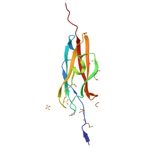
| Immunogen | Recombinant fusion protein containing a sequence corresponding to amino acids 1-85 of human NBR1 (NP_005890.2) |
| Source | Rabbit |
| Category | Polyclonal Antibodies |
| Application | WB, IHC-P, IF/ICC, ELISA |
| Cross Reactivity | Human, Mouse, Rat |
| SWISS | Q14596 |
| Protein Weight | 107kDa |
| Shipping | Ice bag |
Biological Background: NBR1 Function and Localization
- NBR1 (Next to BRCA1 gene 1 protein), also referred to as cell migration-inducing gene 19 protein or membrane component chromosome 17 surface marker 2, is a ubiquitin-binding autophagy receptor.
- It acts as an autophagy adapter by shuttling ubiquitinated cargo to autophagosomes and promoting the formation of protein aggregates. Related references: PMID:24879152, PMID:34471133, PMID:38169523
- NBR1 is involved in host defense and intracellular homeostasis. Related references: PMID:24692539, PMID:33577621
- It regulates innate immune signaling by modulating type I interferon production and targeting ubiquitinated IRF3 for autophagic degradation. Related references: PMID:35914352
- Subcellularly, NBR1 localizes to the cytoplasm, autophagosomes, lysosomes, and the sarcomeric M line.
- Under oxidative stress, NBR1 stabilizes SQSTM1/p62 and activates the KEAP1-NRF2 antioxidant pathway (by similarity).
- NBR1 also has a non-autophagic role in shuttling IL-12 to late endosomes for secretion (by similarity).
- The protein contains zinc-finger domains, is phosphorylated, and participates in host-virus interactions.
Experimental Guidance and Technical Tips
- The immunogen corresponds to the N-terminal 1–85 amino acids of human NBR1; therefore, the antibody may detect all known isoforms, but validation in the specific sample context is advised.
- For Western blotting, use a 4–20% gradient gel and expect a band at ~107 kDa under reducing conditions; validate with positive controls such as HeLa or HEK293T cell lysates.
- In IHC-P, antigen retrieval with citrate buffer (pH 6.0) is recommended, and we suggest testing on human skeletal muscle or immune tissue sections known to express NBR1.
- For IF/ICC, autophagy induction (e.g., by starvation or rapamycin treatment) can enhance punctate staining; include appropriate autophagy markers as co-stains.
- Given the cross-reactivity with mouse and rat, confirm sequence conservation of the immunogen region (residues 1–85) between human and the species of interest before use.
- For ELISA, the antibody can be used as a capture or detection reagent; pairing with a monoclonal NBR1 antibody may improve sandwich assay performance.
CamelBio: Your One-Stop Sourcing Bridge
CamelBio offers this anti-NBR1 rabbit polyclonal antibody as part of a complete suite of IVD raw materials for autophagy, immunity, and oncology research. As your integrated sourcing partner, we supply validated antibody pairs, optimized recombinant proteins, bulk ancillary reagents, and custom development services to accelerate your diagnostic pipeline from concept to clinical application.
Product Datasheet
Anti-NBR1 Rabbit Polyclonal Antibody for WB, IHC-P, IF/ICC, ELISA - Q14596
REQUEST A QUOTE
Our professional team will reply to you within one business day. Please feel free to contact us!