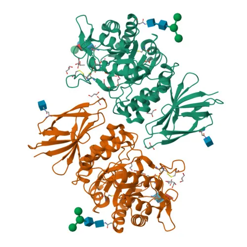
Polyclonal Antibodies
Anti-NAGA Rabbit Polyclonal Antibody for WB, IF/ICC, ELISA - P17050
Item Number : CM0026694
Price varies based on specs and customizations
- Application
- WB, IF/ICC, ELISA
- Cross Reactivity
- Human, Mouse, Rat
- Protein Weight
- 47kDa

Shipping:
Contact us to get shipping details Enjoy On-time Dispatch Guarantee.
Why Choose Us
Easy ordering process, quality products, and dedicated support for your business success.
Core Product Specifications and Parameters
| Parameter | Value |
|---|---|
| Product Name | NAGA Rabbit pAb |
| Remarks/Alias | GALB; D22S674; NAGA |
| Species | Human |
| GeneID (human) | 4668 |
| GeneID | 4668 |
| Immunogen | Recombinant protein|Recombinant fusion protein containing a sequence corresponding to amino acids 312-411 of human NAGA (NP_000253.1). |
| Source | Rabbit |
| Category | Polyclonal Antibodies |
| Application | WB, IF/ICC, ELISA |
| Cross Reactivity | Human, Mouse, Rat |
| SWISS | P17050 |
| Protein Weight | 47kDa |
| Shipping | Ice bag |
Biological Background: Alpha-N-acetylgalactosaminidase (NAGA) Function and Localization
- NAGA encodes alpha-N-acetylgalactosaminidase, also known as alpha-galactosidase B, a lysosomal glycosidase.
- It catalyzes the removal of terminal alpha-N-acetylgalactosamine residues from glycolipids and glycopeptides, essential for glycolipid catabolism.
- The enzyme is localized to the lysosome, where it participates in the degradation of glycoconjugates.
- Post-translational modifications include N-glycosylation (glycoprotein), phosphorylation (phosphoprotein), and the presence of a signal peptide and disulfide bonds, as indicated by UniProt keywords.
- Mutations in NAGA are associated with disease, as noted by the 'Disease variant' keyword, though specific phenotypes may vary; epilepsy is listed among associated keywords.
- The protein has a calculated molecular weight of approximately 46.6 kDa (UniProt), while the antibody product targets a 47 kDa band, consistent with the glycosylated mature form.
Experimental Guidance and Technical Tips
- The immunogen was designed using a recombinant fragment corresponding to amino acids 312–411 near the C-terminus of human NAGA (NP_000253.1). This region may be surface-exposed, potentially enabling recognition of both native and denatured protein.
- For Western blot (WB), a band around 47 kDa is expected; consider using a positive control lysate from tissues or cells known to express NAGA, such as lysosome-rich samples.
- In immunofluorescence/immunocytochemistry (IF/ICC), co-staining with a lysosomal marker is recommended to confirm lysosomal localization.
- The antibody's cross-reactivity with mouse and rat samples suggests conserved epitope recognition; however, sequence alignment is advised to verify epitope conservation if quantitative species comparisons are intended.
- For ELISA applications, optimize coating conditions and use a recombinant NAGA protein or peptide as a standard curve to ensure quantitative accuracy.
CamelBio: Your One-Stop Sourcing Bridge
CamelBio provides diagnostic manufacturers, labs, and research institutes with streamlined access to a wide range of IVD raw materials and services, supporting projects from concept to clinic. For lysosomal enzyme research, such as studies on alpha-N-acetylgalactosaminidase (NAGA) in glycolipid metabolism and related disorders, we offer validated antibody pairs, optimized polyclonal antibodies like anti-NAGA, bulk ancillary reagents, and expert sourcing of rare-target raw materials, ensuring your diagnostic development proceeds with reliability and efficiency.
REQUEST A QUOTE
Our professional team will reply to you within one business day. Please feel free to contact us!