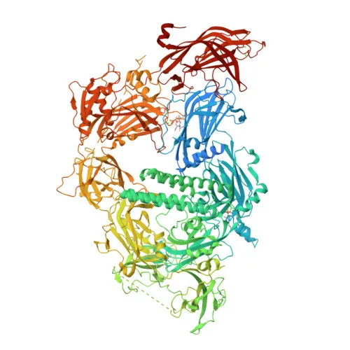
Polyclonal Antibodies
Anti-MYOF Rabbit Polyclonal Antibody (KO Validated) for WB, ELISA - Q9NZM1
Item Number : CM0018172
Price varies based on specs and customizations
- Application
- WB, ELISA
- Cross Reactivity
- Human, Mouse
- Protein Weight
- 235kDa

Shipping:
Contact us to get shipping details Enjoy On-time Dispatch Guarantee.
Why Choose Us
Easy ordering process, quality products, and dedicated support for your business success.
Core Product Specifications and Parameters
| Parameter | Value |
|---|---|
| Product Name | [KO Validated] MYOF Rabbit pAb |
| Remarks/Alias | HAE7; FER1L3; OF |
| Species | Human |
| GeneID (Human) | 26509 |
| GeneID | 26509 |
| Immunogen | Recombinant fusion protein containing a sequence corresponding to amino acids 1-190 of human MYOF (NP_038479.1). |
| Source | Rabbit |
| Category | Polyclonal Antibodies |
| Application | WB, ELISA |
| Cross Reactivity | Human, Mouse |
| SWISS | Q9NZM1 |
| Protein Weight | 235kDa |
| Shipping | Ice bag |
Biological Background: Myoferlin Function and Localization
- Myoferlin (also known as Fer-1-like protein 3) is a calcium/phospholipid-binding protein encoded by the MYOF gene, and is a member of the ferlin family of proteins involved in membrane fusion and repair.
- It plays a critical role in the plasmalemma repair mechanism of endothelial cells, enabling rapid resealing of membranes disrupted by mechanical stress.
- Involved in endocytic recycling and implicated in VEGF signal transduction by regulating the levels of the receptor KDR.
- Localizes to the cell membrane, nucleus membrane, and cytoplasmic vesicle membranes, including late endosome membranes.
- Expressed in myoblast and endothelial cells, with high expression in cardiac and skeletal muscles; also present in lung, and at very low levels in kidney, placenta, and brain.
- Contains multiple C2 domains that mediate calcium-dependent phospholipid binding, and is subject to post-translational modifications including acetylation and phosphorylation.
Experimental Guidance and Technical Tips
The immunogen used for this antibody is a recombinant fusion protein corresponding to the N-terminal region (aa 1-190) of human MYOF. This design may allow recognition of multiple isoforms or splice variants; we recommend verifying specificity in the relevant sample system. For Western blot, note the high molecular weight of myoferlin (~235 kDa); consider using optimized transfer conditions and a suitable membrane for efficient detection. In ELISA assays, titer and working dilution should be empirically determined. As a polyclonal antibody, lot-to-lot consistency may vary; validation of each new lot in your specific application is advisable.
CamelBio: Your One-Stop Sourcing Bridge
CamelBio provides diagnostic manufacturers, labs, and research institutes with one-stop access to IVD raw materials, technical services, and consulting—covering every stage from concept to clinic. Our portfolio includes validated antibody pairs, optimized monoclonal/polyclonal antibodies, bulk ancillary reagents, and rare target raw-material sourcing to support your myoferlin research and assay development needs. With a focus on membrane repair and muscle biology, CamelBio is your partner for reliable, high-quality reagents.
Product Datasheet
Anti-MYOF Rabbit Polyclonal Antibody (KO Validated) for WB, ELISA - Q9NZM1
REQUEST A QUOTE
Our professional team will reply to you within one business day. Please feel free to contact us!