Polyclonal Antibodies
Anti-MYO10 Polyclonal Antibody for WB, IF/ICC, ELISA - Q9HD67
Item Number : CM0015915
Price varies based on specs and customizations
- Application
- WB, IF/ICC, ELISA
- Cross Reactivity
- Human, Mouse
- Protein Weight
- 237kDa

Shipping:
Contact us to get shipping details Enjoy On-time Dispatch Guarantee.
Why Choose Us
Easy ordering process, quality products, and dedicated support for your business success.
Core Product Specifications and Parameters
| Parameter | Value |
|---|---|
| Product Name | MYO10 Rabbit pAb |
| Remarks/Alias | MyoX; MYO10 |
| Species | Human |
| GeneID (Human) | 4651 |
| GeneID | 4651 |
| Immunogen | Recombinant fusion protein containing a sequence corresponding to amino acids 845-944 of human MYO10 (NP_036466.2) |
| Source | Rabbit |
| Category | Polyclonal Antibodies |
| Application | WB, IF/ICC, ELISA |
| Cross Reactivity | Human, Mouse |
| SWISS | Q9HD67 |
| Protein Weight | 237kDa |
| Shipping | Ice bag |
Biological Background: MYO10 Function and Localization
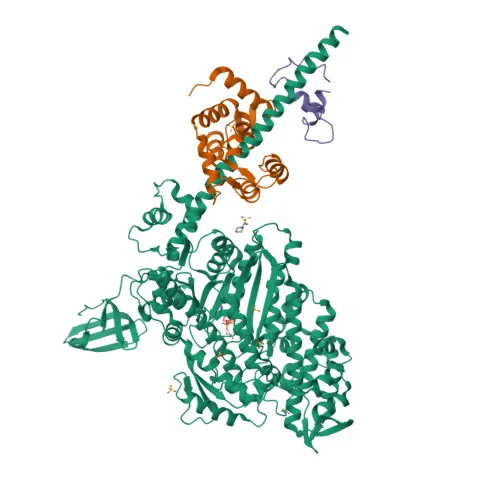
- MYO10, also known as unconventional myosin-X or myosin-10, is an actin-based motor protein with ATPase activity. It binds to actin filaments and bundles, functioning as a plus end-directed motor with enhanced velocity on actin bundles (PubMed:27580874).
Related references:
PMID:27580874 - Its tail domain binds to membranous compartments containing phosphatidylinositol 3,4,5-trisphosphate (PIP3) or integrins, mediating cargo transport along actin filaments. This interaction regulates cell shape, spreading, and adhesion.
- MYO10 stimulates filopodia formation and elongation. In hippocampal neurons, it induces dendritic filopodia by trafficking VASP to filopodial tips, promoting actin elongation.
- An alternative isoform (isoform 2) functions as a dominant-negative regulator, suppressing filopodia-inducing and axon outgrowth-promoting activities of isoform 1, and instead promoting spine formation and spine head expansion in hippocampal neurons.
- Subcellular localization includes cytoplasm, cytosol, cell projections such as lamellipodia, ruffles, and filopodial tips, as well as the cell cortex and filopodial membrane.
- The protein undergoes post-translational modifications including acetylation and phosphorylation. It contains multiple domains: a coiled-coil region, calmodulin-binding motifs, and a pleckstrin homology (PH) domain.
- MYO10 is ubiquitously expressed across tissues, consistent with its fundamental role in cell morphology and migration.
- Keywords associated with MYO10 include: 3D-structure, Actin-binding, Alternative splicing, ATP-binding, Calmodulin-binding, Motor protein, Myosin, Nucleotide-binding, Phosphoprotein, Transport.
Experimental Guidance and Technical Tips
- The immunogen corresponds to a sequence in the tail domain of MYO10 (residues 845–944), which is present in both isoform 1 and likely isoform 2. The antibody may recognize both isoforms, but validation in the relevant sample system is recommended.
- For Western blotting, a band of approximately 237 kDa is expected. Use an appropriate protein ladder for high-molecular-weight proteins and optimize transfer conditions.
- In immunofluorescence/immunocytochemistry, MYO10 localizes to filopodia tips, lamellipodia, and ruffles. Validate fixation and permeabilization protocols to preserve delicate cell projections.
- For ELISA, ensure that the coating antigen is compatible with the immunogen region. Cross-reactivity with mouse is reported, supporting its use in mouse-derived samples.
CamelBio: Your One-Stop Sourcing Bridge
CamelBio provides diagnostic manufacturers and research labs with one-stop access to IVD raw materials, including validated antibodies like this anti-MYO10 polyclonal antibody for cytoskeleton and cell motility studies. Our comprehensive portfolio features antibody pairs, optimized monoclonal/polyclonal antibodies, bulk ancillary reagents, and customized rare target sourcing, helping you move seamlessly from concept to clinic.
REQUEST A QUOTE
Our professional team will reply to you within one business day. Please feel free to contact us!