Monoclonal Antibodies
Anti-MYH7/β-MHC Rabbit PolymAb® Monoclonal Antibody for WB, IF-P, ELISA - P12883
Item Number : CM0007099
Price varies based on specs and customizations
- Application
- WB, IF-P, ELISA
- Cross Reactivity
- Human, Mouse, Rat
- Protein Weight
- 223kDa

Shipping:
Contact us to get shipping details Enjoy On-time Dispatch Guarantee.
Why Choose Us
Easy ordering process, quality products, and dedicated support for your business success.
Core Product Specifications and Parameters
| Parameter | Value |
|---|---|
| Product Name | MYH7/β-MHC Rabbit PolymAb® |
| Remarks/Alias | CMH1; MPD1; SPMD; SPMM; CMD1S; MYHCB; CMYP7A; CMYP7B |
| Species | Human |
| Gene ID | 4625 |
| Immunogen | A synthetic peptide corresponding to a sequence within amino acids 1240-1340 of human MYH7/β-MHC (NP_000248.2) |
| Source | Rabbit |
| Category | Monoclonal Antibodies |
| Application | WB, IF-P, ELISA |
| Cross Reactivity | Human, Mouse, Rat |
| SWISS | P12883 |
| Protein Weight | 223kDa |
| Shipping | Ice bag |
Biological Background: MYH7/β-MHC Function and Localization
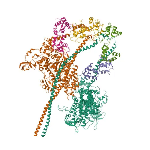
- MYH7, also known as myosin heavy chain 7, MyHC-beta, or MyHC-slow, is the cardiac muscle beta isoform of myosin heavy chain, a major component of the thick filament in sarcomeres.
- As an actin-based motor protein, MYH7 possesses ATPase activity and converts chemical energy into mechanical force, driving muscle contraction in both cardiac and skeletal muscles.
- The protein forms bipolar thick filaments that, together with actin thin filaments, constitute the fundamental contractile units of striated muscle.
- MYH7 is localized predominantly in the cytoplasm, specifically within myofibrils and sarcomeres, where it participates in the sliding filament mechanism of contraction.
- Post-translational modifications such as phosphorylation and methylation have been identified on MYH7 and are thought to regulate its function and interaction with other sarcomeric proteins.
- Mutations in the MYH7 gene are associated with various cardiomyopathies, including hypertrophic and dilated cardiomyopathy, highlighting its critical role in cardiac muscle function.
- At the protein level, MYH7 is detected in both cardiac and skeletal muscle tissues, consistent with its expression in type I slow-twitch fibers.
Experimental Guidance and Technical Tips
- For Western blotting, given the large protein size (~223 kDa), use low-percentage acrylamide gels (e.g., 6-8%) and extended transfer times, and consider wet transfer methods for efficient blotting.
- In immunofluorescence-paraffin (IF-P) experiments, optimize antigen retrieval protocols and detergent permeabilization to ensure antibody penetration into densely packed myofibrillar structures.
- The immunogen peptide lies in the C-terminal region (aa 1240-1340), which includes coiled-coil domains; verify that the sequence is conserved across your species of interest (human, mouse, rat) for optimal cross-reactivity.
- For ELISA-based assays, the synthetic peptide immunogen suggests the antibody may perform well against denatured or peptide targets; consider checking cross-reactivity with related MYH isoforms if nonspecific binding is a concern.
CamelBio: Your One-Stop Sourcing Bridge
CamelBio supports diagnostic manufacturers, core laboratories, and research institutes with comprehensive access to high-quality IVD raw materials. This anti-MYH7/β-MHC antibody supports research into cardiac muscle disorders and sarcomere biology, and CamelBio can assist in sourcing validated antibody pairs, ancillary reagents, and bulk orders for large-scale assay development. From rare targets like MYH7 to routine diagnostics, our supply chain bridges the gap from concept to clinical application.
Product Datasheet
Anti-MYH7/β-MHC Rabbit PolymAb® Monoclonal Antibody for WB, IF-P, ELISA - P12883
REQUEST A QUOTE
Our professional team will reply to you within one business day. Please feel free to contact us!