Monoclonal Antibodies
Anti-Mouse CD8a Monoclonal Antibody (APC) for Flow Cytometry - P01731
Item Number : CM0004917
Price varies based on specs and customizations
- Application
- FC
- Cross Reactivity
- Mouse
- Protein Weight
- 27kDa

Shipping:
Contact us to get shipping details Enjoy On-time Dispatch Guarantee.
Why Choose Us
Easy ordering process, quality products, and dedicated support for your business success.
Core Product Specifications and Parameters
| Parameter | Value |
|---|---|
| Product Name | APC Rabbit anti-Mouse CD8a mAb |
| Remarks/Alias | Ly-2; Ly-B; Ly-35; Lyt-2 |
| Species | Mouse |
| GeneID (Human) | 925 |
| GeneID | 12525 |
| Immunogen | Recombinant protein / Recombinant fusion protein containing a sequence corresponding to amino acids 28-196 of mouse CD8a (NP_001074579.1) |
| Source | Rabbit |
| Category | Monoclonal Antibodies |
| Application | FC |
| Cross Reactivity | Mouse |
| SWISS | P01731 |
| Protein Weight | 27kDa |
| Shipping | Ice bag |
Biological Background: CD8a Function and Localization
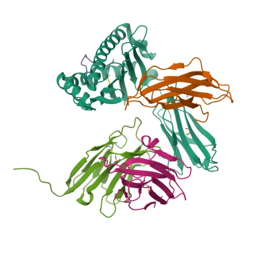
- CD8a (Lyt-2) is an integral membrane glycoprotein that serves as a coreceptor for MHC class I molecules on T cells, facilitating the recognition of peptide antigens derived from cytosolic proteins.
- It interacts simultaneously with the T-cell receptor (TCR) and MHC class I–peptide complexes on antigen-presenting cells (APCs), enhancing T-cell activation.
- CD8a recruits the Src kinase LCK to the TCR-CD3 complex, initiating intracellular signaling cascades that lead to lymphokine production, motility, adhesion, and cytotoxic T-lymphocyte (CTL) activation.
- This coreceptor function enables CTLs to specifically recognize and eliminate virally infected or transformed cells.
- On natural killer (NK) cells, CD8A homodimers contribute to target cell conjugation and lysis, and also support the survival and differentiation of activated lymphocytes into memory CD8⁺ T cells.
- The protein is localized at the cell membrane and contains a single transmembrane helix, with extracellular immunoglobulin-like domains stabilized by disulfide bonds.
- Post-translational modifications include N-linked glycosylation and palmitoylation, which may influence membrane microdomain localization and signaling.
- Alternative splicing generates multiple CD8a isoforms, and the protein is a member of the immunoglobulin superfamily involved in adaptive immunity.
Experimental Guidance and Technical Tips
- The immunogen covers the extracellular region (aa 28–196) of mouse CD8a, suggesting that the antibody likely recognizes the native cell surface form of the protein.
- As a direct APC conjugate, this antibody is ready-to-use for flow cytometry (FC) without the need for secondary detection reagents; consider titrating the optimal antibody concentration for your assay.
- Validate the staining pattern and specificity in your mouse model or cell line of interest, and include appropriate isotype controls to confirm specificity.
CamelBio: Your One-Stop Sourcing Bridge
CamelBio streamlines IVD development by providing a comprehensive catalog of immunodiagnostic antibodies, including this anti-mouse CD8a monoclonal antibody (APC) for flow cytometry. We support diagnostic manufacturers, labs, and research institutes with optimized raw materials, from antibody pairs to bulk ancillary reagents, and offer custom sourcing for rare targets. For your T-cell and immuno-oncology assay needs, count on CamelBio as your dedicated supply partner.
Product Datasheet
Anti-Mouse CD8a Monoclonal Antibody (APC) for Flow Cytometry - P01731
REQUEST A QUOTE
Our professional team will reply to you within one business day. Please feel free to contact us!