Monoclonal Antibodies
Anti-Mouse CD272/BTLA Monoclonal Antibody () for Flow Cytometry - Q7TSA3
Item Number : CM0010875
Price varies based on specs and customizations
- Application
- FC
- Cross Reactivity
- Mouse
- Protein Weight
- 34kDa

Shipping:
Contact us to get shipping details Enjoy On-time Dispatch Guarantee.
Why Choose Us
Easy ordering process, quality products, and dedicated support for your business success.
Core Product Specifications and Parameters
| Parameter | Value |
|---|---|
| Product Name | Rabbit anti-Mouse CD272/BTLA mAb |
| Remarks/Alias | A630002H24 |
| Species | Mouse |
| Gene ID (Human) | 151888 |
| Gene ID | 208154 |
| Immunogen | Recombinant fusion protein containing a sequence corresponding to amino acids 30-176 of mouse CD272/BTLA (NP_001032808.2) |
| Source | Rabbit |
| Category | Monoclonal Antibodies |
| Application | FC |
| Cross Reactivity | Mouse |
| SWISS | Q7TSA3 |
| Protein Weight | 34kDa |
| Shipping | Ice bag |
Biological Background: CD272/BTLA Function and Localization
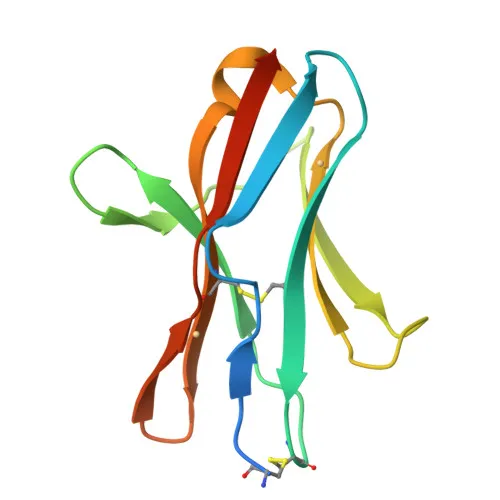
- CD272 (BTLA) is a B- and T-lymphocyte attenuator, an inhibitory receptor belonging to the immunoglobulin superfamily. It contains an extracellular immunoglobulin domain, a transmembrane helix, and a cytoplasmic tail with immunoreceptor tyrosine-based inhibitory motifs (ITIMs).
- BTLA negatively regulates antigen receptor signaling in lymphocytes by recruiting protein tyrosine phosphatases SHP-1 and SHP-2. Related references: PMID:12796776 PMID:14652006
- It interacts with TNFRSF14 (HVEM) in cis (on the same cell) or in trans (between cells). Cis interactions maintain a resting state in naive T cells, while trans interactions provide survival signals to effector T cells during adaptive immune responses. Related references: PMID:19915044
- Expressed in splenic T- and B-cells, lymph node tissues, and also in macrophages, NK cells, and dendritic cells. A polymorphic tissue distribution is observed between different mouse strains.
- The protein is located at the cell membrane as a single-pass type I membrane protein, with an N-terminal signal peptide cleaved during processing.
- Post-translational modifications include glycosylation and phosphorylation, and its structure contains disulfide bonds important for immunoglobulin domain stability.
- Alternative splicing generates multiple isoforms, though the functional significance in mouse is not fully characterized.
- Broadly involved in adaptive immunity, with three-dimensional structural data available.
Experimental Guidance and Technical Tips
- The antibody is directly conjugated to , a red fluorescent dye, suited for flow cytometric detection. Consider titrating the antibody to determine the optimal staining concentration for your experimental system.
- Generated against the extracellular domain (amino acids 30-176), it likely recognizes the native cell surface receptor. Include appropriate isotype controls to ensure specificity.
- For staining immune cells, combine with lineage markers to gate on specific populations (e.g., T cells, B cells, NK cells).
- Standard flow cytometry staining protocols (e.g., 30 min at 4°C) are applicable, but validate fixation steps as they may affect epitope recognition.
- As a rabbit monoclonal antibody, it can be detected with an anti-rabbit IgG secondary reagent if needed, though direct conjugation eliminates this step.
CamelBio: Your One-Stop Sourcing Bridge
CamelBio provides diagnostic manufacturers, labs, and research institutes with a comprehensive range of IVD raw materials and services. This Rabbit anti-Mouse CD272/BTLA monoclonal antibody supports immunology and oncology research, enabling precise detection of BTLA by flow cytometry. Beyond high-performance antibodies, CamelBio offers validated antibody pairs, optimized monoclonal/polyclonal antibodies, bulk ancillary reagents, and rare target raw-material sourcing to streamline your development pipeline from concept to clinic.
Product Datasheet
Anti-Mouse CD272/BTLA Monoclonal Antibody () for Flow Cytometry - Q7TSA3
REQUEST A QUOTE
Our professional team will reply to you within one business day. Please feel free to contact us!