Polyclonal Antibodies
Anti-MLKL Rabbit Polyclonal Antibody for WB, IF-P, IHC-P, ELISA - Q9D2Y4
Item Number : CM0026889
Price varies based on specs and customizations
- Application
- WB, IF-P, IHC-P, ELISA
- Cross Reactivity
- Human, Mouse
- Protein Weight
- 54kDa

Shipping:
Contact us to get shipping details Enjoy On-time Dispatch Guarantee.
Why Choose Us
Easy ordering process, quality products, and dedicated support for your business success.
Core Product Specifications and Parameters
| Parameter | Value |
|---|---|
| Product Name | MLKL Rabbit pAb |
| Remarks/Alias | 9130019I15Rik; MLKL |
| Species | Mouse |
| Gene ID (Human) | 197259 |
| Gene ID | 74568 |
| Immunogen | Recombinant fusion protein containing a sequence corresponding to amino acids 1-53 of mouse MLKL (Q9D2Y4). |
| Source | Rabbit |
| Category | Polyclonal Antibodies |
| Application | WB, IF-P, IHC-P, ELISA |
| Cross Reactivity | Human, Mouse |
| SWISS | Q9D2Y4 |
| Protein Weight | 54kDa |
| Shipping | Ice bag |
Biological Background: MLKL Function and Localization
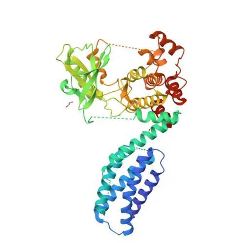
- MLKL (mixed lineage kinase domain-like protein) is a pseudokinase that lacks intrinsic protein kinase activity. Related references: PMID:24012422
- It acts as a critical executor of TNF-induced necroptosis, becoming activated upon RIPK3-mediated phosphorylation, which triggers homotrimerization and translocation to the plasma membrane. Related references: PMID:23835476 PMID:24012422 PMID:24019532
- In response to orthomyxovirus infection, MLKL participates in nuclear necroptosis following ZBP1 sensing of Z-RNA; nuclear RIPK3 phosphorylates MLKL, leading to nuclear envelope disruption and cytosolic DNA leakage. Related references: PMID:32200799 PMID:32296175
- Binding to highly phosphorylated inositol phosphates such as InsP6 is essential for its necroptotic function.
- Subcellular localization spans the cytoplasm, cell membrane, and nucleus, reflecting its dynamic trafficking during cell death signaling.
- It is highly expressed in thymus, colon, intestine, liver, spleen, and lung, with lower levels in skeletal muscle, heart, and kidney; absent in brain.
- Structural features include a coiled-coil domain and a nucleotide-binding region, and it exists in multiple isoforms via alternative splicing.
- MLKL is regulated by phosphorylation, a key post-translational modification for its activation.
Experimental Guidance and Technical Tips
- The immunogen spans the N-terminal region (aa 1-53) of mouse MLKL, which may detect full-length and cleavage products; validate in your relevant sample system.
- For Western blot (WB), the predicted molecular weight is approximately 54 kDa; consider using reducing conditions and appropriate molecular weight markers.
- In immunostaining applications (IF-P, IHC-P), optimize permeabilization and antigen retrieval due to MLKL"s membrane and nuclear localization.
- For ELISA, this polyclonal antibody can serve as either capture or detection reagent; pairing with monoclonal antibodies may improve assay specificity.
- As a polyclonal antibody, lot-to-lot variability may occur; include appropriate controls in each experiment.
CamelBio: Your One-Stop Sourcing Bridge
CamelBio empowers diagnostic manufacturers and research teams with end-to-end access to IVD raw materials, streamlining your journey from concept to clinic. This anti-MLKL antibody supports necroptosis and cell death research, and is just one example of our extensive portfolio covering validated antibody pairs, optimized monoclonal/polyclonal antibodies, bulk ancillary reagents, and specialized raw-material sourcing for rare targets. Partner with us to secure reliable, performance-driven reagents tailored to your assay development needs.
Product Datasheet
Anti-MLKL Rabbit Polyclonal Antibody for WB, IF-P, IHC-P, ELISA - Q9D2Y4
REQUEST A QUOTE
Our professional team will reply to you within one business day. Please feel free to contact us!