Monoclonal Antibodies
Anti-MLKL Monoclonal Antibody for WB, IHC-P, ELISA - Q8NB16
Item Number : CM0010885
Price varies based on specs and customizations
- Application
- WB, IHC-P, ELISA
- Cross Reactivity
- Human, Mouse
- Protein Weight
- 54 kDa

Shipping:
Contact us to get shipping details Enjoy On-time Dispatch Guarantee.
Why Choose Us
Easy ordering process, quality products, and dedicated support for your business success.
Core Product Specifications and Parameters
| Parameter | Value |
|---|---|
| Product Name | [KO Validated] MLKL Mouse mAb |
| Remarks/Alias | hMLKL |
| Species | Human |
| GeneID (human) | 197259 |
| GeneID | 197259 |
| Immunogen | Synthetic peptide: A synthesized peptide derived from human MLKL. |
| Source | Mouse |
| Category | Monoclonal Antibodies |
| Application | WB, IHC-P, ELISA |
| Cross Reactivity | Human, Mouse |
| SWISS | Q8NB16 |
| Protein Weight | 54 kDa |
| Shipping | Ice bag |
Biological Background: MLKL Function and Localization
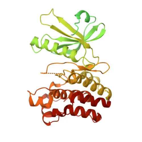
- MLKL (Mixed lineage kinase domain-like protein) is a pseudokinase that plays a critical role in tumor necrosis factor (TNF)-induced necroptosis, a programmed cell death pathway distinct from apoptosis. Related references: PMID:22265413, PMID:22265414, PMID:22421439
- It lacks intrinsic protein kinase activity and functions downstream of RIPK3. Upon phosphorylation by RIPK3, MLKL undergoes homotrimerization and translocates to the plasma membrane, inducing calcium influx and membrane damage to execute necroptotic cell death. Related references: PMID:22265413, PMID:22265414, PMID:22421439
- MLKL also mediates nuclear necroptosis triggered by viral infections. Following activation by ZBP1, nuclear RIPK3 phosphorylates MLKL, leading to disruption of the nuclear envelope and leakage of cellular DNA into the cytosol.
- The protein binds highly phosphorylated inositol phosphates, such as inositol hexakisphosphate (InsP6), which is essential for its necroptotic function. Related references: PMID:29883610
- MLKL localizes to the cytoplasm, cell membrane, and nucleus, consistent with its dynamic role in signaling and cell death execution.
- Alternative splicing generates multiple MLKL isoforms, potentially contributing to regulatory diversity.
- Keywords associated with MLKL include ATP-binding, coiled coil, necrosis, phosphoprotein, and nucleotide-binding, reflecting its structural domains and role in necroptosis signaling.
Experimental Guidance and Technical Tips
- The antibody was raised against a synthetic peptide from human MLKL; it may recognize both denatured and native forms, supporting applications such as Western blotting (WB) and ELISA.
- In Western blot, a band at approximately 54 kDa is expected for both human and mouse MLKL; include appropriate positive controls to confirm specificity.
- For immunohistochemistry (IHC-P), antigen retrieval conditions may require optimization, and validation using known positive tissue sections is recommended.
- The antibody's cross-reactivity with mouse MLKL enables its use in mouse models of necroptosis; however, performance should be validated in the relevant experimental system.
- As MLKL is a phosphoprotein, consider the role of phosphorylation in your experimental context, as activation-induced modifications may affect detection.
- For necroptosis assays, inducing MLKL activation typically requires combined treatments (e.g., TNF-α, Smac mimetic, and a caspase inhibitor); confirm activation status with appropriate controls.
CamelBio: Your One-Stop Sourcing Bridge
As a dedicated IVD raw material supplier, CamelBio offers comprehensive sourcing solutions for antibodies like Anti-MLKL monoclonal antibody, catering to necroptosis and cell death research. From validated antibody pairs and bulk ancillary reagents to rare target sourcing, we support seamless development of immunoassays and diagnostic kits. Partner with us to streamline your workflow from concept to clinic.
REQUEST A QUOTE
Our professional team will reply to you within one business day. Please feel free to contact us!