Polyclonal Antibodies
Anti-MIB1/DIP1 Rabbit Polyclonal Antibody for WB, ELISA - Q86YT6
Item Number : CM0025234
Price varies based on specs and customizations
- Application
- WB, ELISA
- Cross Reactivity
- Human, Mouse, Rat
- Protein Weight
- 110kDa

Shipping:
Contact us to get shipping details Enjoy On-time Dispatch Guarantee.
Why Choose Us
Easy ordering process, quality products, and dedicated support for your business success.
Core Product Specifications and Parameters
| Parameter | Value |
|---|---|
| Product Name | MIB1/DIP1 Rabbit pAb |
| Remarks/Alias | MIB; DIP1; ZZZ6; DIP-1; LVNC7; ZZANK2; MIB1/DIP1 |
| Species | Human |
| Gene ID (Human) | 57534 |
| Gene ID | 57534 |
| Immunogen | Recombinant fusion protein containing a sequence corresponding to amino acids 560-860 of human MIB1/DIP1 (NP_065825.1). |
| Source | Rabbit |
| Category | Polyclonal Antibodies |
| Application | WB, ELISA |
| Cross Reactivity | Human, Mouse, Rat |
| SWISS | Q86YT6 |
| Protein Weight | 110kDa |
| Shipping | Ice bag |
Biological Background: MIB1/DIP1 Function and Localization
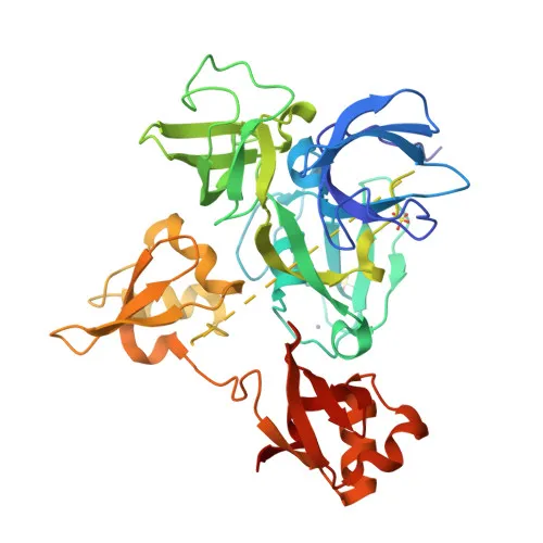
- E3 ubiquitin-protein ligase MIB1 (Mind bomb homolog 1), also known as DAPK-interacting protein 1 (DIP-1) and zinc finger ZZ type with ankyrin repeat domain protein 2, is a member of the RING-type E3 ubiquitin transferase family.
- Positively regulates Delta-mediated Notch signaling by ubiquitinating the intracellular domain of Delta receptors, leading to their endocytosis.
- Mediates ubiquitination and probable proteasomal degradation of DAPK1, thereby antagonizing DAPK1's anti-apoptotic effects to promote TNF-induced apoptosis (by similarity).
- Ubiquitinates centriolar satellite proteins CEP131, CEP290, and PCM1, inhibiting primary cilium formation in proliferating cells.
- Mediates 'Lys-63'-linked polyubiquitination of TBK1, which likely participates in kinase activation.
- During adenovirus infection, mediates ubiquitination of the core-capsid bridging protein, facilitating viral genome delivery into the nucleus.
- Found in the cytoplasm, at the centrosome and centriolar satellites, and at the cell membrane.
- Widely expressed at low levels, with higher expression in the spinal cord, ovary, and brain.
Experimental Guidance and Technical Tips
- The immunogen corresponds to a central region of human MIB1 (aa 560–860); this region shares high sequence identity with mouse and rat MIB1, consistent with the reported cross-reactivity, but experimental verification in the relevant species is recommended.
- For Western blotting, the expected band is at ~110 kDa; consider using 8% SDS-PAGE and a PVDF membrane with standard transfer conditions, and validate with appropriate controls.
- In ELISA applications, the antibody may be used as capture or detection reagent; titrate to determine optimal working concentration for your specific assay format.
CamelBio: Your One-Stop Sourcing Bridge
CamelBio supports diagnostic manufacturers and research laboratories with a comprehensive portfolio of IVD raw materials. For targets like MIB1/DIP1, involved in critical processes such as Notch signaling and apoptosis, CamelBio offers validated polyclonal and monoclonal antibodies, recombinant proteins, and ancillary reagents to streamline your assay development. Whether you need bulk quantities or specialized sourcing for rare targets, CamelBio is your reliable partner from concept to clinic.
REQUEST A QUOTE
Our professional team will reply to you within one business day. Please feel free to contact us!