Monoclonal Antibodies
Anti-MHC Class II (I-A/I-E) Monoclonal Antibody for Flow Cytometry - P18468 / P06342 / Q3U060 / P14435
Item Number : CM0011387
Price varies based on specs and customizations
- Application
- FC
- Cross Reactivity
- Mouse
- Protein Weight
- 30kDa/32kDa/26kDa

Shipping:
Contact us to get shipping details Enjoy On-time Dispatch Guarantee.
Why Choose Us
Easy ordering process, quality products, and dedicated support for your business success.
Core Product Specifications and Parameters
| Parameter | Value |
|---|---|
| Product Name | Rat anti-Mouse MHC Class II (I-A/I-E) mAb |
| Remarks/Alias | Eb; Ia4; H2Eb; Ia-4; H-2Eb; IAb; Ia2; Ia-2; Abeta; H-2Ab; H2-Ab; Rmcs1; I-Abeta; Ia5; Ia-5; H-2Eb2; Iebeta2; A130038H09Rik; Ia1; H2Aa; Ia-1; H-2Aa; Aalpha; IAalpha; I-Aalpha |
| Species | Mouse |
| Gene ID | 14969/14961/381091/14960 |
| Immunogen | Recombinant protein of mouse MHC Class II (I-A/I-E) |
| Source | Rat |
| Category | Monoclonal Antibodies |
| Application | FC |
| Cross Reactivity | Mouse |
| SWISS | P18468/P06342/Q3U060/P14435 |
| Protein Weight | 30kDa/32kDa/26kDa |
| Shipping | Ice bag |
Biological Background: MHC Class II (I-A/I-E) Function and Localization
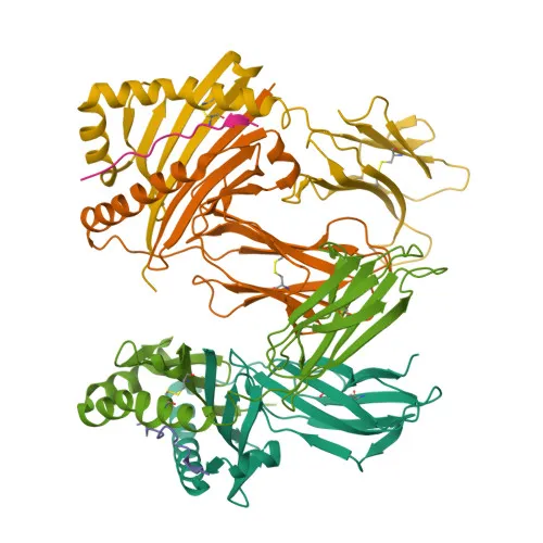
- MHC class II molecules are heterodimeric glycoproteins composed of alpha and beta chains, presenting extracellular peptides to CD4+ T cells. The I-A and I-E isotypes are the classical MHC II molecules in mouse.
- The I-A beta chain (encoded by H2-Ab1) and I-E beta chain (encoded by H2-Eb1) are highly polymorphic membrane proteins that form complexes with their respective alpha chains, I-A alpha and I-E alpha.
- These molecules are critical for adaptive immunity, mediating antigen presentation to T-helper cells and shaping T-cell repertoire during thymic selection.
- Subcellular localization: Predominantly expressed on the plasma membrane of professional antigen-presenting cells (dendritic cells, B cells, macrophages) and thymic epithelial cells.
- Post-translational modifications include disulfide bond formation, N-linked glycosylation, and ubiquitination, which regulate MHC II trafficking and turnover.
- Keywords: 3D-structure, Adaptive immunity, Disulfide bond, Glycoprotein, Immunity, Membrane, MHC II, Reference proteome, Signal, Transmembrane, Transmembrane helix, Ubl conjugation.
Experimental Guidance and Technical Tips
- For flow cytometry, titrate the antibody to determine the optimal staining concentration for your specific cell type and experimental conditions.
- Include appropriate isotype control and viability dye to discriminate dead cells, which may exhibit non-specific binding.
- Validate antibody performance in relevant mouse strains; note that some allotype-specific variations may affect recognition.
- Perform surface staining on live, unfixed cells; if fixation is required, test for potential epitope masking.
- Use fluorochrome-matched compensation controls when performing multicolor flow cytometry.
CamelBio: Your One-Stop Sourcing Bridge
CamelBio offers a comprehensive range of IVD raw materials and sourcing solutions, including validated antibodies, bulk ancillary reagents, and custom procurement services. As a one-stop partner for diagnostic manufacturers and research labs, we can support your MHC Class II and immunology-related projects from concept to clinic. Contact us to explore how our supply chain capabilities can accelerate your development timelines.
Product Datasheet
Anti-MHC Class II (I-A/I-E) Monoclonal Antibody for Flow Cytometry - P18468 / P06342 / Q3U060 / P14435
REQUEST A QUOTE
Our professional team will reply to you within one business day. Please feel free to contact us!