Polyclonal Antibodies
Anti-MGAT4A Rabbit Polyclonal Antibody for WB, ELISA - Q9UM21
Item Number : CM0017852
Price varies based on specs and customizations
- Application
- WB, ELISA
- Cross Reactivity
- Human
- Protein Weight
- 62kDa

Shipping:
Contact us to get shipping details Enjoy On-time Dispatch Guarantee.
Why Choose Us
Easy ordering process, quality products, and dedicated support for your business success.
Core Product Specifications and Parameters
| Parameter | Value |
|---|---|
| Product Name | MGAT4A Rabbit pAb |
| Remarks/Alias | GNT-IV; GnT-4a; GNT-IVA; MGAT4A |
| Species | Human |
| GeneID (Human) | 11320 |
| GeneID | 11320 |
| Immunogen | Recombinant fusion protein containing a sequence corresponding to amino acids 28-100 of human MGAT4A (NP_036346.1) |
| Source | Rabbit |
| Category | Polyclonal Antibodies |
| Application | WB, ELISA |
| Cross Reactivity | Human |
| SWISS | Q9UM21 |
| Protein Weight | 62kDa |
| Shipping | Ice bag |
Biological Background: MGAT4A Function and Localization
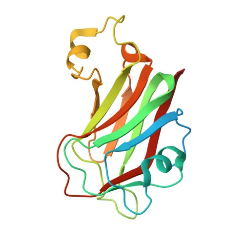
- Alpha-1,3-mannosyl-glycoprotein 4-beta-N-acetylglucosaminyltransferase A (MGAT4A), also known as GNT-IV, GnT-4a, or GNT-IVA, is a key glycosyltransferase encoded by the MGAT4A gene. It belongs to the glycosyltransferase 54 family and undergoes alternative splicing, potentially generating multiple isoforms.
- Catalyzes the transfer of N-acetylglucosamine (GlcNAc) from UDP-GlcNAc to the GlcNAcβ1-2Manα1-3 arm of N-glycan cores via a β1-4 linkage, promoting the synthesis of tri- and tetra-antennary N-linked oligosaccharides.
Related references: PMID:17006639 - Plays a role in glucose homeostasis by mediating glycosylation of the glucose transporter SLC2A2/GLUT2, thereby regulating its cell-surface expression in pancreatic beta cells.
- Predominantly localized to the Golgi apparatus membrane, with a fraction also secreted into the extracellular space, consistent with its role in glycan processing.
- Tissue distribution includes broad expression in pancreas, spleen, thymus, prostate, small intestine, and leukocytes; notably overexpressed in choriocarcinoma cell lines but down-regulated in pancreatic cancer.
- Annotated as a metal-binding, phosphoprotein, and single-pass type II membrane protein (signal-anchor), requiring divalent cations such as Mn²⁺ for enzymatic activity.
Experimental Guidance and Technical Tips
- The immunogen corresponds to residues 28–100 of human MGAT4A (NP_036346.1), an N-terminal region that may help differentiate isoforms. For Western blot, anticipate a band near 62 kDa under reducing conditions; verify using appropriate molecular weight markers.
- Validate antibody performance in relevant cell lysates: consider using choriocarcinoma cell lines (e.g., JEG-3) as positive controls due to reported overexpression, or pancreatic cancer lines where expression is down-regulated for contrast.
- For ELISA, the antibody may be used for direct or indirect detection of MGAT4A, but optimal working dilutions must be determined empirically.
- Because MGAT4A is involved in SLC2A2 glycosylation, this antibody may serve as a tool in studies of glucose transport and diabetes research; ensure appropriate model systems are used.
CamelBio: Your One-Stop Sourcing Bridge
CamelBio provides diagnostic manufacturers, labs, and research institutes with one-stop access to IVD raw materials, technical services, and consulting—covering every stage from concept to clinic. This anti-MGAT4A polyclonal antibody exemplifies our commitment to supplying high-quality reagents for glycosylation and metabolic disease research. Beyond this product, we offer validated antibody pairs, optimized monoclonal antibodies, and bulk ancillary reagents to support your entire assay development pipeline.
REQUEST A QUOTE
Our professional team will reply to you within one business day. Please feel free to contact us!