Monoclonal Antibodies
Anti-MET Rabbit Monoclonal Antibody for WB and ELISA - P08581
Item Number : CM0006287
Price varies based on specs and customizations
- Application
- WB, ELISA
- Cross Reactivity
- Mouse
- Protein Weight
- 156kDa

Shipping:
Contact us to get shipping details Enjoy On-time Dispatch Guarantee.
Why Choose Us
Easy ordering process, quality products, and dedicated support for your business success.
Core Product Specifications and Parameters
| Parameter | Value |
|---|---|
| Product Name | MET Rabbit mAb |
| Remarks/Alias | DA11; HGFR; AUTS9; RCCP2; c-Met; DFNB97; MET |
| Species | Human |
| Gene ID | 4233 |
| Immunogen | Recombinant fusion protein containing a sequence corresponding to amino acids 960-1161 of human MET (NP_000236.2) |
| Source | Rabbit |
| Category | Monoclonal Antibodies |
| Application | WB, ELISA |
| Cross Reactivity | Mouse |
| SWISS | P08581 |
| Protein Weight | 156kDa |
| Shipping | Ice bag |
Biological Background: MET Function and Localization
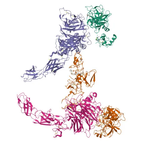
- MET, also known as hepatocyte growth factor receptor (HGFR) or c-Met, is a receptor tyrosine kinase that transduces signals from the extracellular matrix upon binding its ligand HGF.
- Ligand-induced activation triggers autophosphorylation and recruitment of downstream effectors (e.g., PI3K, PLCγ, GRB2, STAT3), leading to activation of RAS-ERK, PI3K-AKT, and PLCγ-PKC pathways that coordinate proliferation, scattering, morphogenesis, and survival.
- During embryogenesis, MET signaling is essential for gastrulation, neuronal precursor migration, angiogenesis, and kidney formation; postnatally it supports wound healing, organ regeneration, and tissue remodeling.
- Subcellularly, MET is primarily located at the membrane (with a secreted form reported) and is expressed in hepatocytes, gastrointestinal epithelia, basal keratinocytes, thyroid, kidney, and brain.
- Tissue expression data show high levels in liver, gastrointestinal tract, thyroid, and kidney (PubMed:26637977). Related references: PMID:26637977
- MET acts as a receptor for Listeria monocytogenes internalin InlB, facilitating host cell entry.
- Post-translational modifications include phosphorylation, glycosylation, and ubiquitination; clinically, MET mutations, alternative splicing, and rearrangements are linked to deafness and multiple cancers.
Experimental Guidance and Technical Tips
- The immunogen spans the cytoplasmic domain (aa 960–1161); the antibody may recognize both full-length and cleaved C-terminal fragments. Consider this when analyzing WB band patterns.
- Standard reducing SDS-PAGE is recommended; non-reducing conditions may be evaluated for native conformation studies.
- For ELISA, use recombinant MET or HGF-stimulated cell lysates to confirm binding; cross-reactivity with mouse MET enables translational studies in mouse models, but validate in the relevant sample system.
- Optimal working dilutions should be determined empirically for each experimental setup.
CamelBio: Your One-Stop Sourcing Bridge
CamelBio provides diagnostic manufacturers and research institutions with streamlined access to high-quality IVD raw materials—from validated antibody pairs and optimized monoclonal antibodies to bulk ancillary reagents and rare target sourcing. MET is a central biomarker in cancer diagnostics and targeted therapy development; we support your MET-related projects with reliable, ready-to-use raw materials and flexible supply solutions. Reach out to explore how our integrated sourcing platform can accelerate your assay development from concept to clinic.
REQUEST A QUOTE
Our professional team will reply to you within one business day. Please feel free to contact us!