Polyclonal Antibodies
Anti-MET Polyclonal Antibody for WB, IHC-P, ELISA - P08581
Item Number : CM0022844
Price varies based on specs and customizations
- Application
- WB, IHC-P, ELISA
- Cross Reactivity
- Human, Mouse, Rat
- Protein Weight
- 156kDa

Shipping:
Contact us to get shipping details Enjoy On-time Dispatch Guarantee.
Why Choose Us
Easy ordering process, quality products, and dedicated support for your business success.
Core Product Specifications and Parameters
| Parameter | Value |
|---|---|
| Product Name | MET Rabbit pAb |
| Remarks/Alias | DA11; HGFR; AUTS9; RCCP2; c-Met; DFNB97; MET |
| Species | Human |
| Gene ID (Human) | 4233 |
| Gene ID | 4233 |
| Immunogen | Recombinant fusion protein containing a sequence corresponding to amino acids 518-840 of human MET (NP_000236.2) |
| Source | Rabbit |
| Category | Polyclonal Antibodies |
| Application | WB, IHC-P, ELISA |
| Cross Reactivity | Human, Mouse, Rat |
| SWISS | P08581 |
| Protein Weight | 156kDa |
| Shipping | Ice bag |
Biological Background: MET Function and Localization
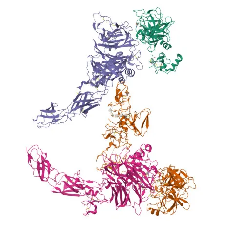
- Hepatocyte growth factor receptor (MET), also known as c-Met, is a receptor tyrosine kinase that transduces signals from HGF ligand to regulate proliferation, scattering, morphogenesis, and survival.
- Ligand binding induces autophosphorylation and recruitment of adaptors (GAB1, GRB2) and effectors (PIK3R1, PLCG1, SRC, STAT3), activating RAS-ERK (morphogenesis) and PI3K-AKT (prosurvival) signaling cascades.
- During embryonic development, MET is essential for gastrulation, neuronal precursor migration, angiogenesis, kidney formation, and skeletal muscle progenitor migration/proliferation (by similarity).
- In adults, MET participates in wound healing, organ regeneration, tissue remodeling, and hematopoietic cell differentiation; may also regulate cortical bone osteogenesis (by similarity).
- Acts as a receptor for Listeria monocytogenes internalin InlB, facilitating bacterial entry into host cells.
- Subcellular localization is primarily at the cell membrane and can be secreted; the mature receptor comprises disulfide-linked α and β subunits.
- Expressed in normal hepatocytes, gastrointestinal epithelial cells, skin keratinocytes, and at high levels in liver, thyroid, kidney, and brain; also detected in metaphyseal bone (PubMed:26637977). Related references: PMID:26637977
- Post-translational modifications include glycosylation, phosphorylation, and ubiquitination, as indicated by UniProt keywords.
Experimental Guidance and Technical Tips
- The immunogen covers amino acids 518–840 of the extracellular domain, likely recognizing native and cleaved MET forms; validate in relevant cell or tissue systems.
- For western blotting, a band around 156 kDa (precursor) or 145 kDa (mature β-chain) is expected under reducing conditions; use appropriate positive controls (e.g., HepG2, A549 lysates).
- In immunohistochemistry (paraffin), optimize antigen retrieval; strong staining is expected in liver and epithelial tissues; confirm specificity with isotype controls.
- Cross-reactivity is stated for human, mouse, and rat; if using other species, pilot testing is advised. As a polyclonal antibody, lot-to-lot consistency should be assessed.
CamelBio: Your One-Stop Sourcing Bridge
CamelBio supports diagnostic manufacturers and research teams with comprehensive access to IVD raw materials, including validated antibody pairs, optimized polyclonal and monoclonal antibodies, and bulk ancillary reagents. We specialize in rare-target sourcing and offer integrated technical services from concept to clinic. For your MET-related projects, we can provide matched antibody pairs, recombinant proteins, and custom development to accelerate assay design.
REQUEST A QUOTE
Our professional team will reply to you within one business day. Please feel free to contact us!