Polyclonal Antibodies
Anti-MELK Rabbit Polyclonal Antibody for WB and ELISA - Q14680
Item Number : CM0013597
Price varies based on specs and customizations
- Application
- WB, ELISA
- Cross Reactivity
- Human, Mouse, Rat
- Protein Weight
- 75kDa

Shipping:
Contact us to get shipping details Enjoy On-time Dispatch Guarantee.
Why Choose Us
Easy ordering process, quality products, and dedicated support for your business success.
Core Product Specifications and Parameters
| Parameter | Value |
|---|---|
| Product Name | MELK Rabbit pAb |
| Remarks/Alias | HPK38; MELK |
| Species | Human |
| GeneID (human) | 9833 |
| GeneID | 9833 |
| Immunogen | Recombinant fusion protein containing a sequence corresponding to amino acids 412-651 of human MELK (NP_055606.1) |
| Source | Rabbit |
| Category | Polyclonal Antibodies |
| Application | WB, ELISA |
| Cross Reactivity | Human, Mouse, Rat |
| SWISS | Q14680 |
| Protein Weight | 75kDa |
| Shipping | Ice bag |
Biological Background: MELK Function and Localization
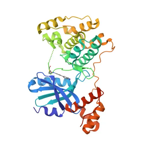
- Maternal embryonic leucine zipper kinase (MELK), also known as pEg3 kinase or hPK38, is a serine/threonine-protein kinase encoded by the MELK gene in humans. It belongs to a broad family of kinases involved in cell cycle regulation and apoptosis.
- MELK acts as an activator of apoptosis by phosphorylating and activating MAP3K5/ASK1, thereby promoting programmed cell death. It also phosphorylates BCL2L14, inhibiting its pro-apoptotic function, which may influence mammary carcinogenesis.
- As a key cell cycle regulator, MELK mediates phosphorylation of CDC25B, promoting its localization to centrosomes and spindle poles during mitosis, which is essential for proper cell division.
- MELK is required for the proliferation of embryonic and postnatal multipotent neural progenitors, highlighting its role in stem cell self-renewal and neurogenesis.
- The kinase also participates in splicing regulation by phosphorylating ZNF622 during mitosis, inhibiting spliceosome assembly and redirecting ZNF622 to the nucleus.
- Subcellular localization studies indicate MELK is present at the cell membrane, although its kinase activity is critical in cytoplasmic and nuclear compartments depending on the cellular context.
- Tissue expression profiling shows MELK is prominently expressed in placenta, kidney, thymus, testis, ovary, and intestine, with potential roles in primitive hematopoiesis.
- Post-translational modifications and structural features include ATP-binding, calcium- and lipid-binding domains, and multiple phosphorylation sites as cataloged in the Phosphoprotein keyword entry.
Experimental Guidance and Technical Tips
- The immunogen corresponds to amino acids 412–651 of human MELK, a region that may provide target specificity; verify sequence alignment across species if cross-reactivity with mouse or rat is critical.
- For Western blot, a detection of a band at approximately 75 kDa is expected. Consider using dilution ranges such as 1:500–1:2000 with appropriate positive controls (e.g., cell lines with known MELK expression) and validate in your sample system.
- In ELISA, performance may depend on target protein conformation and coating conditions; consider using recombinant MELK protein or cell lysates to determine optimal antibody dilutions.
- Because cross-reactivity is reported for human, mouse, and rat, tissue-specific expression patterns should be taken into account when selecting positive controls.
CamelBio: Your One-Stop Sourcing Bridge
As a dedicated IVD raw-material partner, CamelBio offers validated antibody pairs, optimized monoclonal and polyclonal antibodies, bulk ancillary reagents, and rare target sourcing—covering your needs from concept to clinic. For research focusing on cell cycle, apoptosis, or stem cell biology targets like MELK, CamelBio provides one-stop access to high-quality antibodies and technical support to accelerate your diagnostic development.
REQUEST A QUOTE
Our professional team will reply to you within one business day. Please feel free to contact us!