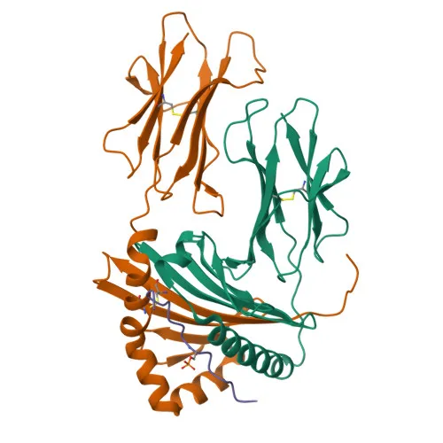
Monoclonal Antibodies
Anti-Melan-A/MART-1 Rabbit Monoclonal Antibody for WB, IF, IHC, ELISA - Q16655
Item Number : CM0007214
Price varies based on specs and customizations
- Application
- WB, IF/ICC, IHC-P, ELISA
- Cross Reactivity
- Human
- Protein Weight
- 13kDa

Shipping:
Contact us to get shipping details Enjoy On-time Dispatch Guarantee.
Why Choose Us
Easy ordering process, quality products, and dedicated support for your business success.
Core Product Specifications and Parameters
| Parameter | Value |
|---|---|
| Product Name | Melan-A/MART-1 Rabbit mAb |
| Remarks/Alias | MART1; MART-1; Melan-A/MART-1 |
| Species | Human |
| GeneID (human) | 2315 |
| GeneID | 2315 |
| Immunogen | Synthetic peptide corresponding to a sequence within amino acids 11-110 of human MLANA (NP_005502.1) |
| Source | Rabbit |
| Category | Monoclonal Antibody |
| Application | WB, IF/ICC, IHC-P, ELISA |
| Cross Reactivity | Human |
| SWISS | Q16655 |
| Protein Weight | 13kDa |
| Shipping | Ice bag |
Biological Background: Melan-A/MART-1 Function and Localization
- Melan-A, also designated MART-1 (Melanoma Antigen Recognized by T-cells 1), is a melanocyte lineage-specific protein encoded by the MLANA gene.
- It functions as a key regulator of melanosome biogenesis – it stabilizes the G protein-coupled receptor GPR143 and is essential for the correct expression, trafficking, and processing of the melanocyte protein PMEL (Pmel17/silver), a structural component of stage II melanosomes.
- The protein localizes to the endoplasmic reticulum membrane, Golgi apparatus, trans-Golgi network, and nascent melanosomes, consistent with its role in intracellular sorting.
- Expression of Melan-A/MART-1 is normally restricted to melanocytes, the retinal pigment epithelium, and melanoma cells, making it a valuable diagnostic marker for melanocytic lesions.
- The UniProt annotation highlights post-translational phosphorylation (a phosphoprotein) and a single-pass type III transmembrane topology.
Experimental Guidance and Technical Tips
- The rabbit monoclonal antibody was raised against a synthetic peptide spanning amino acids 11–110 of human MLANA. This N-terminal region is predicted to be cytoplasmic; verify epitope accessibility in your fixation/permeabilization protocol for IF/IHC.
- In Western blotting, the predicted molecular weight of Melan-A is ~13 kDa. Use high-resolution gradient gels and appropriate low-molecular-weight markers. For reliable detection, confirm the band pattern with a known positive control such as melanoma cell lysate (e.g., A375, SK-MEL-28).
- For immunocytochemistry and immunohistochemistry, both frozen and formalin-fixed paraffin-embedded samples may be tested. Antigen retrieval (e.g., citrate buffer pH 6) can enhance staining in FFPE sections.
- Cross-reactivity is reported for human only; validate specificity in the relevant sample system if non-human models are needed.
CamelBio: Your One-Stop Sourcing Bridge
CamelBio serves as a one-stop sourcing bridge for IVD raw materials, connecting diagnostic manufacturers and researchers with validated antibody pairs, optimized monoclonal and polyclonal antibodies, and bulk ancillary reagents. If you are developing assays for melanoma marker detection or melanocyte biology, our supply chain can support the procurement of this anti-Melan-A/MART-1 antibody together with complementary raw materials, from concept to clinical application.
Product Datasheet
Anti-Melan-A/MART-1 Rabbit Monoclonal Antibody for WB, IF, IHC, ELISA - Q16655
REQUEST A QUOTE
Our professional team will reply to you within one business day. Please feel free to contact us!