Polyclonal Antibodies
Anti-MBNL1 Rabbit Polyclonal Antibody for WB - Q9NR56
Item Number : CM0030804
Price varies based on specs and customizations
- Application
- WB, IHC-P, IF/ICC, ELISA
- Cross Reactivity
- Human, Mouse, Rat
- Protein Weight
- 42kDa

Shipping:
Contact us to get shipping details Enjoy On-time Dispatch Guarantee.
Why Choose Us
Easy ordering process, quality products, and dedicated support for your business success.
Core Product Specifications and Parameters
| Parameter | Value |
|---|---|
| Product Name | MBNL1 Rabbit pAb |
| Remarks/Alias | EXP; MBNL; MBNL1 |
| Species | Human |
| GeneID (Human) | 4154 |
| GeneID | 4154 |
| Immunogen | Recombinant fusion protein containing a sequence corresponding to amino acids 1-382 of human MBNL1 (NP_997176.1) |
| Source | Rabbit |
| Category | Polyclonal Antibodies |
| Application | WB, IHC-P, IF/ICC, ELISA |
| Cross Reactivity | Human, Mouse, Rat |
| SWISS | Q9NR56 |
| Protein Weight | 42kDa |
| Shipping | Ice bag |
Biological Background: MBNL1 Function and Localization
- MBNL1 (Muscleblind-like protein 1, also known as Triplet-expansion RNA-binding protein) is an RNA-binding protein that mediates pre-mRNA alternative splicing regulation.
- It acts as either an activator or repressor of splicing on specific pre-mRNA targets; for example, it inhibits cardiac troponin-T (TNNT2) exon inclusion but induces insulin receptor (IR) exon inclusion in muscle, and antagonizes CELF protein splicing activity.
- MBNL1 binds to the consensus sequence 5'-YGCU(U/G)Y-3' and to stem-loop structures within target RNAs, including expanded CUG repeat RNA associated with myotonic dystrophy.
- Together with RNA-binding proteins RBPMS and RBFOX2, MBNL1 activates vascular smooth muscle cell alternative splicing events (PubMed:37548402).
Related references: PMID:37548402 - Subcellularly, MBNL1 localizes to the nucleus, cytoplasm, and cytoplasmic granules.
- It is highly expressed in cardiac and skeletal muscle, as well as during myoblast differentiation, with lower protein-level expression in other tissues including heart, brain, placenta, lung, liver, kidney, and pancreas.
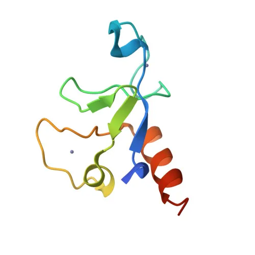
- MBNL1 is a zinc-finger phosphoprotein; post-translational modifications, including phosphorylation, may regulate its activity and localization.
Experimental Guidance and Technical Tips
- The immunogen covers amino acids 1-382 of human MBNL1, a region encompassing the CCCH-type zinc-finger RNA-binding domains; this design likely targets conserved functional epitopes important for RNA binding.
- For Western blot (WB), test a range of antibody dilutions on lysates from muscle tissues (e.g., cardiac or skeletal muscle) where MBNL1 is enriched; the predicted band is around 42 kDa, but validate actual migration in your system.
- Immunohistochemistry (IHC-P) and immunofluorescence (IF/ICC) may require optimization of antigen retrieval and permeabilization; use positive and negative control tissues to confirm specificity.
- For ELISA, assess binding to recombinant MBNL1 protein or peptide and optimize coating and detection conditions for your assay.
- The antibody is reported to cross-react with mouse and rat; however, always validate reactivity in the relevant species before quantitative applications.
CamelBio: Your One-Stop Sourcing Bridge
CamelBio provides diagnostic manufacturers, laboratories, and research institutes with comprehensive access to IVD raw materials, technical services, and consulting, supporting every stage from concept to clinic. For RNA splicing and muscle biology research, we offer high-quality MBNL1 antibodies alongside validated antibody pairs, optimized monoclonal/polyclonal antibodies, and bulk ancillary reagents. Our sourcing expertise extends to rare targets, helping streamline your molecular diagnostics development.
REQUEST A QUOTE
Our professional team will reply to you within one business day. Please feel free to contact us!