Monoclonal Antibodies
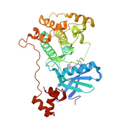
Anti-MARK2 Rabbit Monoclonal Antibody for WB and ELISA - Q7KZI7
Item Number : CM0008334
Price varies based on specs and customizations
- Application
- WB, ELISA
- Cross Reactivity
- Human, Mouse, Rat
- Protein Weight
- 88kDa

Shipping:
Contact us to get shipping details Enjoy On-time Dispatch Guarantee.
Why Choose Us
Easy ordering process, quality products, and dedicated support for your business success.
Core Product Specifications and Parameters
| Parameter | Value |
|---|---|
| Product Name | MARK2 Rabbit mAb |
| Remarks/Alias | EMK1; EMK-1; PAR-1; Par1b; Par-1b; MARK2 |
| Species | Human |
| Gene ID | 2011 |
| Immunogen | Synthetic Peptide corresponding to a sequence within amino acids 689-788 of human MARK2 (Q7KZI7) |
| Source | Rabbit |
| Category | Monoclonal Antibodies |
| Application | WB, ELISA |
| Cross Reactivity | Human, Mouse, Rat |
| SWISS | Q7KZI7 |
| Protein Weight | 88kDa |
| Shipping | Ice bag |
Biological Background: MARK2 Function and Localization
- MARK2 (also known as EMK-1, PAR-1, Par1b) is a serine/threonine-protein kinase that phosphorylates the microtubule-associated protein TAU (MAPT) at KXGS motifs, promoting TAU detachment from microtubules and microtubule disassembly, thereby regulating cell polarity.
Related references: PMID:23666762 - It plays a broad role in cell polarity by phosphorylating additional microtubule-associated proteins MAP2 and MAP4 at similar motifs, leading to their dissociation from the cytoskeleton.
- MARK2 regulates neuronal polarization and migration through phosphorylation of DCX and KIF13B, influencing 14-3-3 protein interactions and microtubule-dependent transport processes.
- It modulates Wnt signaling by phosphorylating disheveled proteins (DVL1, DVL2, DVL3), acting as a positive regulator of the pathway.
- Through phosphorylation of HDAC7, MARK2 promotes its interaction with 14-3-3 and nuclear export, thereby regulating histone deacetylase function.
- In response to ER stress or starvation-induced autophagy, MARK2 phosphorylates UBAC2 at Ser-223, facilitating ER-phagy.
Related references: PMID:39284914 - Subcellular localization includes the cell membrane, cytoplasm, lateral cell membrane, cytoskeleton, and dendritic projections, consistent with its roles in polarity and trafficking.
- High expression in heart, brain, skeletal muscle, and pancreas, with lower expression in lung, liver, and kidney, aligns with its physiological functions.
Experimental Guidance and Technical Tips
- The immunogen is a synthetic peptide from the C-terminal region (aa 689-788) of human MARK2, which shares high sequence identity with mouse and rat orthologs, supporting the observed cross-reactivity.
- For Western blot applications, the predicted molecular weight is approximately 88 kDa; we recommend using positive control lysates from cell lines with known MARK2 expression (e.g., HeLa, HEK293) and optimizing antibody dilution.
- In ELISA assays, consider using recombinant MARK2 protein or the immunogen peptide as the capture antigen; validate assay conditions with a standard curve to ensure linearity and sensitivity.
- Due to its role in cell polarity and microtubule dynamics, additional functional validation in immunofluorescence or immunohistochemistry may require fixation/permeabilization optimization to preserve the native localization.
CamelBio: Your One-Stop Sourcing Bridge
CamelBio provides diagnostic manufacturers, labs, and research institutes with one-stop access to IVD raw materials, technical services, and consulting—covering every stage from concept to clinic. Our portfolio includes this high-quality anti-MARK2 rabbit monoclonal antibody for cell polarity and neurobiology research, alongside validated antibody pairs, bulk ancillary reagents, and custom antibody development services. From rare target sourcing to large-scale production, we streamline your procurement process to accelerate assay development.
REQUEST A QUOTE
Our professional team will reply to you within one business day. Please feel free to contact us!