Polyclonal Antibodies
Anti-MARK1 Rabbit Polyclonal Antibody for WB, IHC-P, ELISA - Q9P0L2
Item Number : CM0027156
Price varies based on specs and customizations
- Application
- WB, IHC-P, ELISA
- Cross Reactivity
- Human, Mouse
- Protein Weight
- 89kDa

Shipping:
Contact us to get shipping details Enjoy On-time Dispatch Guarantee.
Why Choose Us
Easy ordering process, quality products, and dedicated support for your business success.
Core Product Specifications and Parameters
| Parameter | Value |
|---|---|
| Product Name | MARK1 Rabbit pAb |
| Remarks/Alias | MARK; Par1c; Par-1c; MARK1 |
| Species | Human |
| GeneID (Human) | 4139 |
| GeneID | 4139 |
| Immunogen | Recombinant fusion protein containing a sequence corresponding to amino acids 560-700 of human MARK1 (NP_061120.3) |
| Source | Rabbit |
| Category | Polyclonal Antibodies |
| Application | WB, IHC-P, ELISA |
| Cross Reactivity | Human, Mouse |
| SWISS | Q9P0L2 |
| Protein Weight | 89kDa |
| Shipping | Ice bag |
Biological Background: MARK1 Function and Localization
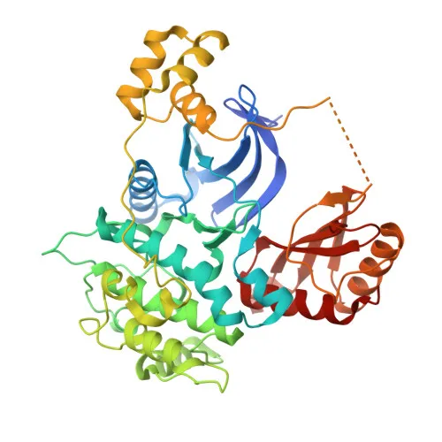
- MARK1 (MAP/microtubule affinity-regulating kinase 1) is a serine/threonine-protein kinase that belongs to the MARK family, which also includes MARK2, MARK3, and MARK4.
- It phosphorylates microtubule-associated proteins such as MAP2, MAP4, and MAPT/TAU at KXGS motifs, leading to their detachment from microtubules and contributing to microtubule disassembly. Related references: PMID:23666762
- MARK1 plays a critical role in regulating cell polarity and neuronal migration by modulating the cytoskeleton, and it phosphorylates DCX (doublecortin) to influence neuronal migration.
- The kinase acts as a positive regulator of the Wnt signaling pathway, likely through phosphorylation of dishevelled proteins (DVL1, DVL2, DVL3).
- Alternative splicing produces multiple isoforms of MARK1, which may have distinct functional roles.
- Subcellularly, MARK1 localizes to the cell membrane, cytoplasm, cytoskeleton, and dendrites, consistent with its role in cell polarity and microtubule dynamics.
- It is highly expressed in the heart, skeletal muscle, brain, fetal brain, and fetal kidney, indicating tissue-specific functions in these organs.
- MARK1 requires magnesium for its kinase activity and ATP binding, and it is regulated by phosphorylation and lipid binding.
Experimental Guidance and Technical Tips
- For Western blotting, anticipate a band near 89 kDa; validate signal using lysates from tissues with high MARK1 expression such as brain or heart.
- In IHC-P, paraffin-embedded human or mouse brain sections may yield specific staining; optimize antigen retrieval conditions and confirm with appropriate controls.
- The polyclonal nature of this antibody may enhance ELISA sensitivity, but evaluate potential cross-reactivity with other MARK family members experimentally.
- Consider using the immunogen sequence (amino acids 560–700) to assess antibody specificity, particularly if full-length protein detection varies.
- When studying MARK1 phosphorylation-dependent signaling, include phosphatase inhibitors in lysis and assay buffers.
CamelBio: Your One-Stop Sourcing Bridge
CamelBio offers diagnostic manufacturers and research labs streamlined access to validated IVD raw materials like this anti-MARK1 polyclonal antibody, supporting projects in kinase signaling and neuroscience. Our comprehensive portfolio includes optimized monoclonal and polyclonal antibodies, bulk ancillary reagents, and custom sourcing for rare targets, ensuring a reliable supply chain from concept to clinic. Partner with us to accelerate your assay development with quality raw materials and expert technical support.
REQUEST A QUOTE
Our professional team will reply to you within one business day. Please feel free to contact us!