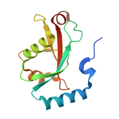
Polyclonal Antibodies
Anti-MAP1LC3B Polyclonal Antibody for WB, ELISA - Q9GZQ8
Item Number : CM0020949
Price varies based on specs and customizations
- Application
- WB, ELISA
- Cross Reactivity
- Human, Rat
- Protein Weight
- 14kDa

Shipping:
Contact us to get shipping details Enjoy On-time Dispatch Guarantee.
Why Choose Us
Easy ordering process, quality products, and dedicated support for your business success.
Core Product Specifications and Parameters
| Parameter | Value |
|---|---|
| Product Name | MAP1LC3B Rabbit pAb |
| Remarks/Alias | LC3B; ATG8F; MAP1LC3B-a; MAP1A/1BLC3; 3B |
| Species | Human |
| Gene ID (Human) | 81631 |
| Immunogen | Synthetic peptide within amino acids 1-50 of human MAP1LC3B (NP_073729.1) |
| Source | Rabbit |
| Category | Polyclonal Antibodies |
| Application | WB, ELISA |
| Cross Reactivity | Human, Rat |
| SWISS | Q9GZQ8 |
| Protein Weight | 14kDa |
| Shipping | Ice bag |
Biological Background: MAP1LC3B Function and Localization
- MAP1LC3B is a ubiquitin-like modifier that conjugates to autophagosomal membranes, promoting elongation of the phagophore membrane during autophagosome formation. Related references: PMID:20418806 PMID:23209295 PMID:28017329
- Plays a critical role in mitophagy by anchoring autophagolysosomes to mitochondrial outer membranes via binding C-18 ceramides, facilitating elimination of damaged mitochondria and regulating mitochondrial quality. Related references: PMID:22922758 PMID:23209295 PMID:28017329
- Involved in selective autophagy pathways, including ER-phagy where it interacts with the reticulophagy receptor TEX264 to remodel ER subdomains into autophagosomes under nutrient stress. Related references: PMID:31006537 PMID:31006538
- Promotes primary ciliogenesis by removing OFD1 from centriolar satellites via the autophagic pathway. Related references: PMID:24089205
- Recruits cofactor JMY to phagophore membranes, stimulating actin nucleation to support autophagosome biogenesis. Related references: PMID:30420355
- Localizes to autophagosome membranes, mitochondrial membranes, and the cytoskeleton, consistent with its role in membrane trafficking and autophagy.
- Most abundant in heart, brain, skeletal muscle, and testis, with low expression in liver.
- Processed and lipidated via a ubiquitin-like conjugation pathway, attaching to phosphatidylethanolamine on forming autophagosomes.
Experimental Guidance and Technical Tips
- The immunogen corresponds to the N-terminal region (aa 1-50) of human MAP1LC3B. For WB, the antibody may detect full-length and potentially N-terminally processed forms; validate cleavage patterns in induced vs. uninduced autophagy samples.
- Under reducing conditions, anticipate a major band at ~14 kDa (LC3-I) and a faster-migrating band corresponding to lipidated LC3-II; consider using autophagy inhibitors (e.g., chloroquine) to distinguish them.
- For ELISA applications, test with recombinant MAP1LC3B protein or peptide as a positive control; assess cross-reactivity with other LC3 family members if specificity is critical.
- Optimize antibody dilution and blocking conditions using tissue or cell lysates from human or rat origin, based on the reported cross-reactivity.
- Inclusion of positive and negative autophagy controls (e.g., rapamycin treatment, ATG5 knockout cells) is recommended to confirm authentic LC3-II detection.
CamelBio: Your One-Stop Sourcing Bridge
CamelBio equips IVD manufacturers and research labs with a streamlined pathway from target discovery to clinical kit development. For autophagy-related targets like MAP1LC3B, we offer validated antibody pairs, optimized polyclonal and monoclonal antibodies, bulk ancillary reagents, and specialized sourcing of rare raw materials. Our integrated supply model ensures consistent, scalable access to critical components for ELISA, WB, and other immunoassay platforms.
REQUEST A QUOTE
Our professional team will reply to you within one business day. Please feel free to contact us!