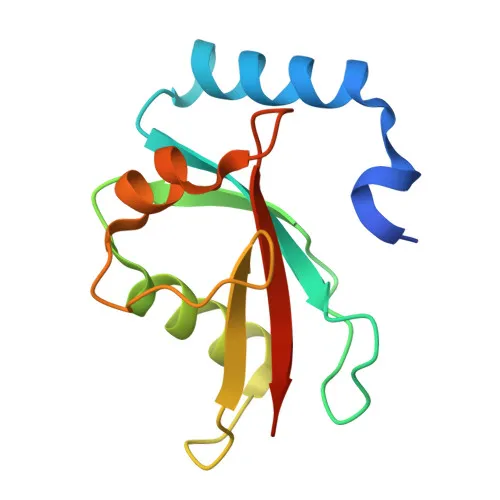
Monoclonal Antibodies
Anti-MAP1LC3A Rabbit Monoclonal Antibody for WB, IP, ELISA - Q9H492
Item Number : CM0002243
Price varies based on specs and customizations
- Application
- WB, IP, ELISA
- Cross Reactivity
- Human, Mouse, Rat
- Protein Weight
- 14kDa

Shipping:
Contact us to get shipping details Enjoy On-time Dispatch Guarantee.
Why Choose Us
Easy ordering process, quality products, and dedicated support for your business success.
Core Product Specifications and Parameters
| Parameter | Value |
|---|---|
| Product Name | MAP1LC3A Rabbit mAb |
| Remarks/Alias | LC3; LC3A; ATG8E; MAP1ALC3; MAP1BLC3; MAP1LC3A |
| Species | Human |
| GeneID (Human) | 84557 |
| GeneID | 84557 |
| Immunogen | Synthetic Peptide|A synthetic peptide corresponding to a sequence within amino acids 1-100 of human MAP1LC3A (Q9H492). |
| Source | Rabbit |
| Category | Monoclonal Antibodies |
| Application | WB, IP, ELISA |
| Cross Reactivity | Human, Mouse, Rat |
| SWISS | Q9H492 |
| Protein Weight | 14kDa |
| Shipping | Ice bag |
Biological Background: MAP1LC3A Function and Localization
- Microtubule-associated protein 1 light chain 3 alpha (MAP1LC3A), also known as LC3, ATG8E, or MAP1ALC3, is a core autophagy-related ubiquitin-like modifier essential for autophagosome formation.
- Plays a key role in the elongation of the phagophore membrane during macroautophagy, distinguishing its function from the GABARAP/GATE-16 subfamily, which acts primarily in autophagosome maturation. Related references: PMID:20713600
- Through interaction with the reticulophagy receptor TEX264, MAP1LC3A participates in remodeling endoplasmic reticulum subdomains into autophagosomes under nutrient stress, facilitating ER turnover via selective autophagy. Related references: PMID:31006537 PMID:31006538
- Localizes to cytoplasmic autophagosome membranes and is also associated with the cytoskeleton and endomembrane system, reflecting its dynamic role in vesicle trafficking and autophagy.
- Tissue expression is most abundant in heart, brain, liver, skeletal muscle, and testis, with absence in thymus and peripheral blood leukocytes, suggesting tissue-specific autophagic activity.
- Classified under keywords including autophagy, Ubl conjugation pathway, and alternative splicing, highlighting its processing and conjugation mechanisms critical for function.
Experimental Guidance and Technical Tips
- The immunogen corresponds to N-terminal residues 1–100; the antibody may recognize both full-length and processed (lipidated) forms of LC3A. Validate detection specificity in your experimental system.
- For Western blot, expect a band near 14–16 kDa; the lipidated LC3-II form migrates faster than the cytosolic LC3-I form. Use lysosomal inhibitors (e.g., chloroquine or bafilomycin A1) to monitor autophagic flux.
- This antibody is suitable for immunoprecipitation and ELISA. Optimize antibody concentrations empirically, and include appropriate controls for each application.
CamelBio: Your One-Stop Sourcing Bridge
CamelBio connects diagnostic manufacturers and research teams with high-quality IVD raw materials, technical services, and consulting support from concept to clinical application. For autophagy-related targets like MAP1LC3A, our sourcing capabilities include validated antibody pairs, optimized monoclonal/polyclonal antibodies, bulk ancillary reagents, and rare target procurement to streamline your development workflow. Let CamelBio be your partner in advancing autophagy research and clinical diagnostics.
REQUEST A QUOTE
Our professional team will reply to you within one business day. Please feel free to contact us!