Polyclonal Antibodies
Anti-MALT1 Rabbit Polyclonal Antibody for WB, IF/ICC, ELISA - Q9UDY8
Item Number : CM0023844
Price varies based on specs and customizations
- Application
- WB, IF/ICC, ELISA
- Cross Reactivity
- Human, Mouse, Rat
- Protein Weight
- 92kDa

Shipping:
Contact us to get shipping details Enjoy On-time Dispatch Guarantee.
Why Choose Us
Easy ordering process, quality products, and dedicated support for your business success.
Core Product Specifications and Parameters
| Parameter | Value |
|---|---|
| Product Name | MALT1 Rabbit pAb |
| Remarks/Alias | MLT; MLT1; IMD12; PCASP1; MALT1 |
| Species | Human |
| GeneID (Human) | 10892 |
| GeneID | 10892 |
| Immunogen | Recombinant fusion protein containing a sequence corresponding to amino acids 125-450 of human MALT1 (NP_776216.1) |
| Source | Rabbit |
| Category | Polyclonal Antibodies |
| Application | WB, IF/ICC, ELISA |
| Cross Reactivity | Human, Mouse, Rat |
| SWISS | Q9UDY8 |
| Protein Weight | 92kDa |
| Shipping | Ice bag |
Biological Background: MALT1 Function and Localization
-
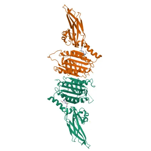
MALT1 (mucosa-associated lymphoid tissue lymphoma translocation protein 1), also known as paracaspase, is a protease that forms CBM (CARD-BCL10-MALT1) complexes with BCL10 and CARD domain-containing proteins (CARD9, CARD11, CARD14) to activate NF-κB and MAP kinase p38 pathways, driving expression of pro-inflammatory cytokines and chemokines. Related references: PMID:11262391, PMID:18264101, PMID:24074955
-
MALT1-dependent cleavage of BCL10 plays a critical role in T-cell antigen receptor-induced integrin adhesion, highlighting its scaffolding and enzymatic functions in immune signaling. Related references: PMID:11262391, PMID:18264101
-
In T cells, MALT1 cleaves N4BP1 following TCR stimulation, leading to inactivation of N4BP1 and modulation of immune responses. Related references: PMID:31133753
-
MALT1 promotes Th17 cell differentiation by cleaving RC3H1 and ZC3H12A, releasing their cooperatively repressed targets.
-
MALT1 may also possess ubiquitin ligase activity by binding to and inducing oligomerization of TRAF6, thereby activating TRAF6 ligase function. Related references: PMID:14695475
-
It localizes predominantly to the cytoplasm, perinuclear region, and nucleus, reflecting its multifunctional roles in signal transduction.
-
MALT1 is highly expressed in peripheral blood mononuclear cells, with lower levels in bone marrow, thymus, lymph node, and very low expression in colon and lung.
-
The protein is subject to acetylation, phosphorylation, and disulfide bond formation, and chromosomal rearrangements involving MALT1 are implicated in MALT lymphoma. Alternative splicing produces multiple isoforms.
Experimental Guidance and Technical Tips
- The immunogen spans amino acids 125–450, encompassing a portion of the immunoglobulin-like domain and part of the paracaspase domain; consider this region’s conservation when evaluating cross-reactivity in mouse and rat samples.
- For Western blotting, the predicted molecular weight is approximately 92 kDa; optimize gel percentage and transfer conditions accordingly for high-molecular-weight proteins.
- MALT1 expression is highest in lymphoid tissues and PBMCs; validate the antibody in the relevant cell or tissue lysate to confirm detection sensitivity.
- For immunofluorescence/ICC, assess subcellular localization patterns (cytoplasmic, perinuclear, or nuclear) in stimulated versus unstimulated cells to capture translocation dynamics.
CamelBio: Your One-Stop Sourcing Bridge
CamelBio offers MALT1 antibodies and related NF-κB pathway reagents as part of a comprehensive portfolio for immunology and oncology research. As a one-stop IVD raw-material supplier, we provide validated antibody pairs, optimized monoclonal and polyclonal antibodies, bulk ancillary reagents, and custom sourcing for rare targets like MALT1—streamlining your assay development from concept to clinic.
REQUEST A QUOTE
Our professional team will reply to you within one business day. Please feel free to contact us!