Polyclonal Antibodies
Anti-MAGT1 Polyclonal Antibody for WB, ELISA - Q9H0U3
Item Number : CM0028427
Price varies based on specs and customizations
- Application
- WB, ELISA
- Cross Reactivity
- Human
- Protein Weight
- 38kDa

Shipping:
Contact us to get shipping details Enjoy On-time Dispatch Guarantee.
Why Choose Us
Easy ordering process, quality products, and dedicated support for your business success.
Core Product Specifications and Parameters
| Parameter | Value |
|---|---|
| Product Name | MAGT1 Rabbit pAb |
| Remarks/Alias | IAP, XMEN, MRX95, OST3B, CDG1CC, PRO0756, SLC58A1, bA217H1.1, MAGT1 |
| Species | Human |
| GeneID (Human) | 84061 |
| Immunogen | Recombinant fusion protein containing a sequence corresponding to amino acids 60-220 of human MAGT1 (NP_115497.4) |
| Source | Rabbit |
| Category | Polyclonal Antibodies |
| Application | WB, ELISA |
| Cross Reactivity | Human |
| SWISS | Q9H0U3 |
| Protein Weight | 38kDa |
| Shipping | Ice bag |
Biological Background: MAGT1 Function and Localization
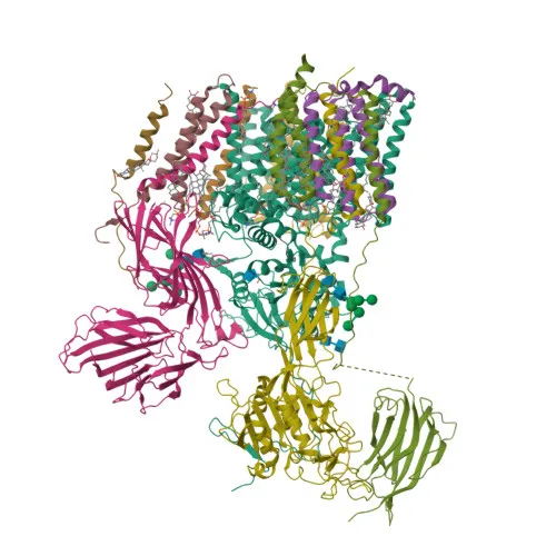
- MAGT1 (also known as Implantation-associated protein IAP or Magnesium transporter protein 1 MagT1) is a subunit of the STT3B-containing form of the oligosaccharyltransferase (OST) complex.
- It acts as an accessory component that catalyzes N-glycosylation of STT3B-dependent substrates, transferring high mannose oligosaccharides to asparagine residues. Related references: PMID:31831667
- The protein is specifically required for the glycosylation of acceptor sites near cysteine residues, functioning redundantly with TUSC3. Its oxidized form may form transient mixed disulfides with substrates.
- MAGT1 also has oxidoreductase-independent roles in the OST complex, possibly involved in substrate recognition.
- It is proposed to indirectly play a role in Mg2+ transport in epithelial cells.
- Subcellularly, MAGT1 localizes to the cell membrane and the endoplasmic reticulum membrane.
- Expression is ubiquitous, with very low levels detected in brain, lung, and kidney.
- MAGT1 is associated with congenital disorders of glycosylation (e.g., CDG1CC) and X-linked immunodeficiency with magnesium defect (XMEN), as reflected by disease-related variants.
Experimental Guidance and Technical Tips
- The immunogen spans amino acids 60–220, a region likely exposed in the luminal/extracellular domain. For Western blot, expect a band at ~38 kDa. Use reducing conditions as the protein contains disulfide bonds.
- Validate the antibody initially with positive control lysates from human cell lines such as HEK293 or HeLa, which express MAGT1.
- For ELISA, the antibody can be used as a capture or detection reagent; optimal working concentrations should be determined by titration.
- Consider pre-absorbing against the fusion partner if non-specific background is observed.
CamelBio: Your One-Stop Sourcing Bridge
CamelBio offers diagnostic manufacturers and research laboratories a seamless supply of validated IVD raw materials, including this anti-MAGT1 polyclonal antibody for glycosylation and membrane protein studies. Beyond antibodies, we provide comprehensive sourcing for optimized monoclonal/polyclonal pairs, bulk ancillary reagents, and rare-target raw materials—streamlining your development from concept to clinic. Partner with us for reliable, one‑stop procurement tailored to your IVD needs.
REQUEST A QUOTE
Our professional team will reply to you within one business day. Please feel free to contact us!