Polyclonal Antibodies
Anti-LYVE1 Rabbit Polyclonal Antibody for WB, ELISA - Q9Y5Y7
Item Number : CM0025601
Price varies based on specs and customizations
- Application
- WB, ELISA
- Cross Reactivity
- Human, Mouse
- Protein Weight
- 35kDa

Shipping:
Contact us to get shipping details Enjoy On-time Dispatch Guarantee.
Why Choose Us
Easy ordering process, quality products, and dedicated support for your business success.
Core Product Specifications and Parameters
| Parameter | Specification |
|---|---|
| Product Name | LYVE1 Rabbit pAb |
| Remarks/Alias | HAR; XLKD1; LYVE-1; CRSBP-1; LYVE1 |
| Species | Human |
| GeneID (Human) | 10894 |
| GeneID | 10894 |
| Immunogen | Recombinant fusion protein containing a sequence corresponding to amino acids 20-238 of human LYVE1 (NP_006682.2) |
| Source | Rabbit |
| Category | Polyclonal Antibodies |
| Application | WB, ELISA |
| Cross Reactivity | Human, Mouse |
| SWISS | Q9Y5Y7 |
| Protein Weight | 35kDa |
| Shipping | Ice bag |
Biological Background: Lymphatic vessel endothelial hyaluronic acid receptor 1 (LYVE1) Function and Localization
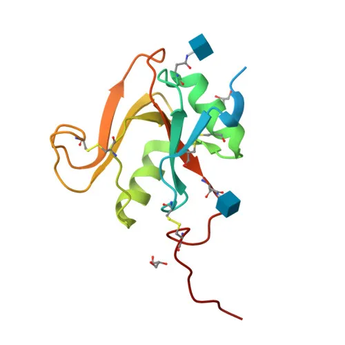
- LYVE1 (Lymphatic Vessel Endothelial Hyaluronic Acid Receptor 1), also known as CRSBP-1 or XLKD1, is a type I transmembrane glycoprotein belonging to the Link protein superfamily.
- It acts as a ligand-specific transporter shuttling between the trans-Golgi network (TGN) and the plasma membrane, and may mediate hyaluronan (HA) uptake for catabolism within lymphatic endothelial cells or transport into lymphatic lumen. Related references: PMID:10037799
- LYVE1 binds pericellular hyaluronan matrices on leukocytes, facilitating cell adhesion and migration through lymphatic endothelium. Related references: PMID:26823460
- It plays a role in autocrine regulation of cell growth mediated by growth regulators containing cell surface retention sequence binding (CRS) motifs.
- The protein is primarily expressed on the surface of endothelial cells lining lymphatic vessels, consistent with its specialized function in the lymphatic system.
- Characteristic structural features include a signal peptide, a single transmembrane helix, disulfide bonds, and N-linked glycosylation sites, typical of membrane receptors.
Experimental Guidance and Technical Tips
- The immunogen corresponds to the extracellular domain (aa 20-238) of human LYVE1, so the antibody likely recognizes native or properly folded protein; denaturing conditions for WB may require optimization.
- For Western blot, expect a band around 35-50 kDa due to glycosylation; use reduced and non-reduced conditions and validate with appropriate positive controls (e.g., lymphatic endothelial cell lysates).
- In ELISA, consider testing against recombinant LYVE1 protein or cell lysates; since it is a polyclonal antibody, direct, capture, or sandwich formats may be feasible.
- Cross-reactivity with mouse extends utility to in vivo models; however, always confirm performance in the specific sample matrix.
- As with any polyclonal antibody, lot-to-lot consistency should be verified, and critical experiments may benefit from additional validation (e.g., blocking with immunogen).
CamelBio: Your One-Stop Sourcing Bridge
CamelBio is your dedicated partner for IVD raw material procurement, offering a comprehensive portfolio that includes this anti-LYVE1 polyclonal antibody as well as validated antibody pairs, recombinant antigens, and ancillary reagents for lymphatic and hyaluronan-related research and diagnostics. Whether you need bulk polyclonal antibodies, custom monoclonal development, or rare target sourcing, our one-stop supply chain ensures continuity and technical support from concept to clinic. Let CamelBio accelerate your IVD assay development with reliable, research-grade materials and flexible supply solutions.
REQUEST A QUOTE
Our professional team will reply to you within one business day. Please feel free to contact us!