Polyclonal Antibodies
Anti-Lyve1 Rabbit Polyclonal Antibody for WB and ELISA - Q8BHC0
Item Number : CM0018907
Price varies based on specs and customizations
- Application
- WB, ELISA
- Cross Reactivity
- Mouse, Rat
- Protein Weight
- 34kDa

Shipping:
Contact us to get shipping details Enjoy On-time Dispatch Guarantee.
Why Choose Us
Easy ordering process, quality products, and dedicated support for your business success.
Core Product Specifications and Parameters
| Parameter | Value |
|---|---|
| Product Name | Lyve1 Rabbit pAb |
| Remarks/Alias | Xlkd1; Lyve-1; Crsbp-1; 1200012G08Rik; Lyve1 |
| Species | Mouse |
| GeneID | 114332 |
| Immunogen | Recombinant fusion protein containing a sequence corresponding to amino acids 30-228 of mouse Lyve1 (NP_444477.2) |
| Source | Rabbit |
| Category | Polyclonal Antibodies |
| Application | WB, ELISA |
| Cross Reactivity | Mouse, Rat |
| SWISS | Q8BHC0 |
| Protein Weight | 34kDa |
| Shipping | Ice bag |
Biological Background: Lyve1 Function and Localization
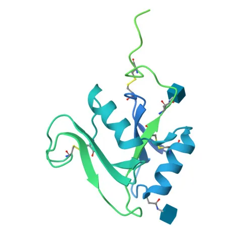
- Lyve1, also known as lymphatic vessel endothelial hyaluronic acid receptor 1, CRSBP-1, or XLKD1, is a transmembrane receptor encoded by the Lyve1 gene in mouse.
- It acts as a ligand-specific transporter between the TGN and plasma membrane, involved in autocrine growth regulation, and may function as a hyaluronan transporter for catabolism within lymphatic endothelial cells or transport into lymph nodes (PubMed:10187853). Related references: PMID:10187853
- Through binding to hyaluronan matrices on leukocytes, Lyve1 promotes cell adhesion and migration across lymphatic endothelium (By similarity).
- Subcellularly, Lyve1 is a membrane protein, consistent with its transport and adhesion functions.
- The protein is characterized as a glycoprotein with a signal peptide, a transmembrane helix, and disulfide bonds, reflecting its structure as a cell surface receptor.
Experimental Guidance and Technical Tips
- The immunogen (amino acids 30–228) corresponds to the extracellular domain, likely enhancing recognition of the native membrane-bound Lyve1; consider validating specificity in target tissue lysates.
- For Western blot, a band around 34 kDa is expected, but glycosylation may increase the apparent molecular weight; we recommend titrating the antibody and including appropriate controls.
- In ELISA applications, this antibody can be used as a capture or detection reagent; validate with recombinant protein or cell lysates.
- Given cross-reactivity with rat, the antibody may be employed in rat models; however, we advise verifying reactivity in the specific experimental system.
CamelBio: Your One-Stop Sourcing Bridge
CamelBio supports diagnostic manufacturers and researchers with a comprehensive portfolio of IVD raw materials, including optimized antibodies for lymphatic and vascular targets like Lyve1. Whether you need validated antibody pairs, bulk ancillary reagents, or assistance in sourcing rare raw materials, our one-stop platform streamlines your development from concept to clinic. Contact us to discuss tailored procurement solutions for your next diagnostic project.
REQUEST A QUOTE
Our professional team will reply to you within one business day. Please feel free to contact us!