Monoclonal Antibodies
Anti-LYVE1 Rabbit Monoclonal Antibody for WB, IF-P, IHC-P, ELISA - Q8BHC0
Item Number : CM0005050
Price varies based on specs and customizations
- Application
- WB, IF-P, IHC-P, ELISA
- Cross Reactivity
- Mouse, Rat
- Protein Weight
- 34kDa

Shipping:
Contact us to get shipping details Enjoy On-time Dispatch Guarantee.
Why Choose Us
Easy ordering process, quality products, and dedicated support for your business success.
Core Product Specifications and Parameters
| Parameter | Value |
|---|---|
| Product Name | LYVE1 Rabbit mAb |
| Remarks/Alias | Xlkd1; Lyve-1; Crsbp-1; 1200012G08Rik; Lyve1 |
| Species | Mouse |
| GeneID (human) | 10894 |
| GeneID | 114332 |
| Immunogen | Recombinant fusion protein containing a sequence corresponding to amino acids 30-228 of mouse Lyve1. (NP_444477.2). |
| Source | Rabbit |
| Category | Monoclonal Antibodies |
| Application | WB, IF-P, IHC-P, ELISA |
| Cross Reactivity | Mouse, Rat |
| SWISS | Q8BHC0 |
| Protein Weight | 34kDa |
| Shipping | Ice bag |
Biological Background: LYVE1 Function and Localization
- LYVE1 is a ligand-specific transporter that mediates trafficking between the trans-Golgi network and the plasma membrane, and plays a role in autocrine regulation of cell growth mediated by growth regulators containing cell surface retention sequence binding (CRS). Related references: PMID:10187853
- It functions as a hyaluronan (HA) transporter, either mediating HA uptake for catabolism within lymphatic endothelial cells or transport into the lumen of afferent lymphatic vessels.
- LYVE1 binds to pericellular hyaluronan matrices deposited on the surface of leukocytes and facilitates cell adhesion and migration through lymphatic endothelium.
- Also known as cell surface retention sequence-binding protein 1 (CRSBP-1) and extracellular link domain-containing protein 1 (XLKD1), reflecting its alternative functions.
- Localized to the membrane, consistent with its roles in transport and cell adhesion.
- Post-translational modifications include glycosylation and disulfide bond formation, and it is a glycoprotein receptor with a signal peptide and a transmembrane helix.
- Mouse LYVE1 has a molecular weight of approximately 34.6kDa (34574 Da).
Experimental Guidance and Technical Tips
- For Western blotting (WB), consider a starting dilution range of 1:500–1:2000 and use protein markers to confirm the ~34kDa band.
- In immunohistochemistry-paraffin (IHC-P) and immunofluorescence-paraffin (IF-P), validate the antibody in lymphatic-rich tissues such as lymph nodes or skin from mouse and rat.
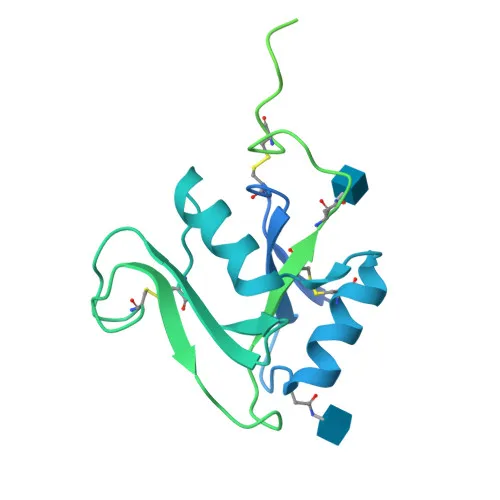
- The immunogen corresponds to the extracellular region (amino acids 30–228), suggesting potential recognition of both native and denatured LYVE1.
- Cross-reactivity with rat is indicated; verify performance in rat samples prior to experimental use.
- For ELISA, optimize coating and detection conditions according to standard protocols for monoclonal antibodies.
CamelBio: Your One-Stop Sourcing Bridge
CamelBio provides diagnostic manufacturers and research institutes with a comprehensive portfolio of IVD raw materials, including this high-quality LYVE1 monoclonal antibody for lymphatic biology and tumor metastasis studies. Our supply capabilities extend to validated antibody pairs, optimized monoclonal/polyclonal antibodies, bulk ancillary reagents, and rare target raw-material sourcing, ensuring a streamlined path from research to clinical application. With expertise from concept to clinic, CamelBio is your trusted partner for all IVD raw-material needs.
Product Datasheet
Anti-LYVE1 Rabbit Monoclonal Antibody for WB, IF-P, IHC-P, ELISA - Q8BHC0
REQUEST A QUOTE
Our professional team will reply to you within one business day. Please feel free to contact us!