Monoclonal Antibodies
Anti-LMAN1 Rabbit Monoclonal Antibody for WB, IHC-P, ELISA - P49257
Item Number : CM0011186
Price varies based on specs and customizations
- Application
- WB, IHC-P, ELISA
- Cross Reactivity
- Human, Mouse, Rat
- Protein Weight
- 54kDa

Shipping:
Contact us to get shipping details Enjoy On-time Dispatch Guarantee.
Why Choose Us
Easy ordering process, quality products, and dedicated support for your business success.
Core Product Specifications and Parameters
| Parameter | Specification |
|---|---|
| Product Name | LMAN1 Rabbit mAb |
| Remarks/Alias | MR60, gp58, F5F8D, FMFD1, MCFD1, ERGIC53, ERGIC-53, LMAN1 |
| Species | Human |
| Gene ID (Human) | 3998 |
| Immunogen | A synthetic peptide corresponding to a sequence within amino acids 1-100 of human LMAN1 (P49257). |
| Source | Rabbit |
| Category | Monoclonal Antibodies |
| Application | WB, IHC-P, ELISA |
| Cross Reactivity | Human, Mouse, Rat |
| SWISS | P49257 |
| Protein Weight | 54kDa |
| Shipping | Ice bag |
Biological Background: LMAN1 Function and Localization
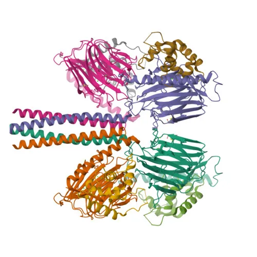
- Protein ERGIC-53 (also known as ER-Golgi intermediate compartment 53 kDa protein, Gp58, and intracellular mannose-specific lectin MR60) is encoded by the LMAN1 gene and functions as a mannose-specific lectin.
- It may recognize sugar residues of glycoproteins, glycolipids, or glycosylphosphatidylinositol anchors, and could be involved in the sorting or recycling of proteins and/or lipids.
- The LMAN1-MCFD2 complex acts as a specific cargo receptor for the ER-to-Golgi transport of selected proteins.
- Subcellularly, LMAN1 localizes to the endoplasmic reticulum-Golgi intermediate compartment (ERGIC) membrane, Golgi apparatus membrane, and endoplasmic reticulum membrane.
- It is ubiquitously expressed across tissues.
- Structural features include a signal peptide, lectin domain, transmembrane helix, disulfide bonds, and sites for phosphorylation and metal binding.
- UniProt keywords highlight its roles in ER-Golgi transport, lectin-mediated recognition, and protein trafficking.
Experimental Guidance and Technical Tips
- The immunogen corresponds to the N-terminal region (aa 1-100) of human LMAN1, encompassing the signal peptide and part of the lectin domain; consider verifying specificity via knockout or knockdown models.
- For Western blot, the glycosylated protein migrates at approximately 54 kDa; include positive lysates from human, mouse, or rat cells as controls.
- In IHC-P, optimization of antigen retrieval and antibody titration is advised; validate staining patterns in LMAN1-expressing tissues.
- For ELISA, pairing with a detection antibody targeting a distinct epitope or using recombinant LMAN1 protein can aid assay development; confirm linearity and sensitivity with standard curves.
CamelBio: Your One-Stop Sourcing Bridge
CamelBio provides comprehensive one-stop sourcing of IVD raw materials, including validated monoclonal antibodies like this LMAN1 rabbit mAb, optimized antibody pairs, and bulk ancillary reagents to accelerate diagnostic development. With expertise in protein trafficking and lectin biology, CamelBio bridges supply gaps for rare targets and supports projects from concept to clinic.
REQUEST A QUOTE
Our professional team will reply to you within one business day. Please feel free to contact us!