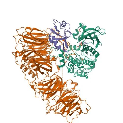
Polyclonal Antibodies
Anti-LLGL1 Rabbit Polyclonal Antibody for WB, IHC-P, IF/ICC, ELISA - Q15334
Item Number : CM0018751
Price varies based on specs and customizations
- Application
- WB, IHC-P, IF/ICC, ELISA
- Cross Reactivity
- Human, Mouse, Rat
- Protein Weight
- 115kDa

Shipping:
Contact us to get shipping details Enjoy On-time Dispatch Guarantee.
Why Choose Us
Easy ordering process, quality products, and dedicated support for your business success.
Core Product Specifications and Parameters
| Parameter | Value |
|---|---|
| Product Name | LLGL1 Rabbit pAb |
| Remarks/Alias | DLG4; HUGL; LLGL; Lgl1; Mgl1; HUGL1; HUGL-1; LLGL1 |
| Species | Human |
| GeneID (Human) | 3996 |
| GeneID | 3996 |
| Immunogen | Recombinant fusion protein containing a sequence corresponding to amino acids 1-190 of human LLGL1 (NP_004131.3) |
| Source | Rabbit |
| Category | Polyclonal Antibodies |
| Application | WB, IHC-P, IF/ICC, ELISA |
| Cross Reactivity | Human, Mouse, Rat |
| SWISS | Q15334 |
| Protein Weight | 115kDa |
| Shipping | Ice bag |
Biological Background: LLGL1 Function and Localization
- LLGL1 (Lethal(2) giant larvae protein homolog 1) is a homolog of the Drosophila tumor suppressor gene l(2)gl and is also known as Hugl-1, DLG4, and HUGL1.
- It functions as a cortical cytoskeleton protein that maintains cell polarity and epithelial integrity by participating in a protein complex at the plasma membrane.
- LLGL1 regulates mitotic spindle orientation, proliferation, differentiation, and tissue organization of neuroepithelial cells, playing a critical role in neurodevelopment.
- It is involved in axonogenesis by activating RAB10, thereby regulating vesicular membrane trafficking toward the axonal plasma membrane.
- The protein localizes to early endosome membranes, Golgi apparatus (including trans-Golgi network), cell projections/axons, and associates with the cytoskeleton.
- Tissue expression is highest in brain, kidney, and muscle, while it is barely detected in heart and placenta; importantly, LLGL1 is downregulated or lost in many tumor cell lines and advanced-stage tumors.
- LLGL1 contains WD repeats and is subject to phosphorylation, with a molecular weight of approximately 115 kDa.
Experimental Guidance and Technical Tips
- The immunogen used to generate this antibody corresponds to the N-terminal region (amino acids 1–190) of human LLGL1; consider verifying specificity by comparing staining patterns in LLGL1-positive vs. knockdown/knockout models.
- For Western blot, the predicted molecular weight of LLGL1 is ~115 kDa; include appropriate molecular weight markers and validate detection in lysates from brain or kidney tissues.
- IHC-P may require optimization of antigen retrieval conditions (e.g., pH6 citrate buffer vs. pH9 EDTA) depending on tissue type and fixation.
- When using IF/ICC, note that LLGL1 associates with membranes and cytoskeletal structures, so permeabilization protocols should be carefully optimized to preserve epitope accessibility.
CamelBio: Your One-Stop Sourcing Bridge
CamelBio provides diagnostic manufacturers and research laboratories with integrated access to IVD raw materials, technical services, and consulting. This anti-LLGL1 antibody supports studies of cell polarity and tumor suppression, areas relevant to neurobiology and oncology. As a one-stop sourcing partner, CamelBio offers validated antibody pairs, bulk ancillary reagents, and customized procurement for challenging targets, helping you streamline development from concept to clinic.
Product Datasheet
Anti-LLGL1 Rabbit Polyclonal Antibody for WB, IHC-P, IF/ICC, ELISA - Q15334
REQUEST A QUOTE
Our professional team will reply to you within one business day. Please feel free to contact us!