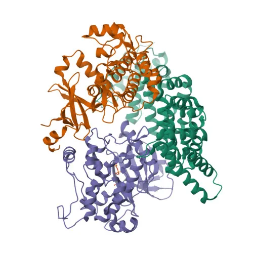
Monoclonal Antibodies
Anti-LKB1 Rabbit Monoclonal Antibody for WB, IHC-P, IP, ELISA - Q15831
Item Number : CM0004747
Price varies based on specs and customizations
- Application
- WB, IHC-P, IP, ELISA
- Cross Reactivity
- Human, Mouse, Rat
- Protein Weight
- 49kDa

Shipping:
Contact us to get shipping details Enjoy On-time Dispatch Guarantee.
Why Choose Us
Easy ordering process, quality products, and dedicated support for your business success.
Core Product Specifications and Parameters
| Parameter | Value |
|---|---|
| Product Name | [KO Validated] LKB1 Rabbit mAb |
| Remarks/Alias | PJS; LKB1; hLKB1; B1 |
| Species | Human |
| Gene ID (Human) | 6794 |
| Gene ID | 6794 |
| Immunogen | Recombinant fusion protein containing a sequence corresponding to amino acids 134-433 of human LKB1. (NP_000446.1). |
| Source | Rabbit |
| Category | Monoclonal Antibodies |
| Application | WB, IHC-P, IP, ELISA |
| Cross Reactivity | Human, Mouse, Rat |
| SWISS | Q15831 |
| Protein Weight | 49kDa |
| Shipping | Ice bag |
Biological Background: LKB1 (STK11) Function and Localization
- LKB1 (STK11) is a serine/threonine kinase and established tumor suppressor, also known as hLKB1, PJS, and renal carcinoma antigen NY-REN-19.
- It serves as the master upstream activator of AMPK and 12 AMPK-related kinases (e.g., BRSK1/2, MARK1-4, NUAK1/2, SIK1-3) by phosphorylating their T-loop residues, thereby regulating energy metabolism, cell polarity, and growth. Related references: PMID:12805220, PMID:14517248, PMID:14976552
- Through AMPK activation, LKB1 controls hepatic glucose homeostasis, autophagy induction during nutrient deprivation, and germinal center B-cell differentiation. Related references: PMID:14976552
- LKB1 is critical for cytoskeletal remodeling and cellular polarity; in neurons, it phosphorylates BRSK1/2 to drive axon initiation and specification. Related references: PMID:15016379, PMID:14976552
- In the DNA damage response, LKB1 interacts with p53/TP53, is recruited to the CDKN1A/WAF1 promoter, and participates in p53-dependent transcription and apoptosis. Related references: PMID:11430832, PMID:17108107, PMID:21317932
- Together with NUAK1, LKB1 phosphorylates CDKN1A after UV irradiation, promoting its degradation to enable efficient DNA repair. Related references: PMID:25329316
- LKB1 dynamically localizes to the nucleus, cytoplasm, mitochondria, and late endosomes, with its membrane association mediated by prenylation and palmitoylation.
- It is ubiquitously expressed with highest levels in testis and fetal liver, and has an additional role in spermiogenesis.
Experimental Guidance and Technical Tips
- The immunogen sequence (aa 134-433) covers the kinase domain and C-terminal regulatory regions; consider performing epitope competition studies to confirm binding specificity.
- For western blot, a band around 49 kDa is expected; validate detection in human, mouse, and rat samples due to reported cross-reactivity.
- For IHC-P, optimize antigen retrieval (e.g., citrate buffer pH 6.0) and include appropriate tissue controls (testis, liver) known to express LKB1 abundantly.
- In IP experiments, verify co-immunoprecipitation of known LKB1 interactors such as STRAD or MO25 under native conditions.
- The antibody's reactivity in ELISA suggests potential for sandwich pair development; however, test with recombinant LKB1 or cell lysates to establish linear range.
CamelBio: Your One-Stop Sourcing Bridge
CamelBio provides diagnostic manufacturers and research labs with comprehensive access to validated antibody pairs, optimized monoclonal and polyclonal antibodies, bulk ancillary reagents, and rare target raw materials. For LKB1 and related tumor suppressors, we offer integrated sourcing solutions to streamline your assay development from concept to clinic. Our one-stop supply model accelerates your IVD projects in kinase signaling and metabolic research areas.
Product Datasheet
Anti-LKB1 Rabbit Monoclonal Antibody for WB, IHC-P, IP, ELISA - Q15831
REQUEST A QUOTE
Our professional team will reply to you within one business day. Please feel free to contact us!