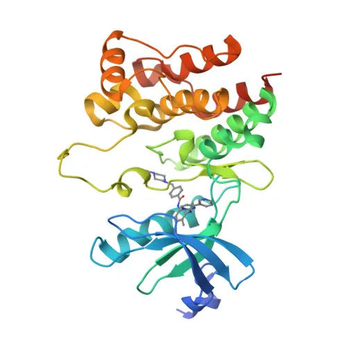
Monoclonal Antibodies
Anti-LCK Rabbit Monoclonal Antibody for WB, IF-P, ELISA - P06239
Item Number : CM0007681
Price varies based on specs and customizations
- Application
- WB, IF-P, ELISA
- Cross Reactivity
- Human, Mouse
- Protein Weight
- 58kDa

Shipping:
Contact us to get shipping details Enjoy On-time Dispatch Guarantee.
Why Choose Us
Easy ordering process, quality products, and dedicated support for your business success.
Core Product Specifications and Parameters
| Parameter | Value |
|---|---|
| Product Name | LCK Rabbit mAb |
| Remarks/Alias | LSK; YT16; IMD22; p56lck; pp58lck; LCK |
| Species | Human |
| GeneID (human) | 3932 |
| GeneID | 3932 |
| Immunogen | Recombinant protein|Recombinant fusion protein containing a sequence corresponding to amino acids 1-210 of human LCK (NP_005347.3). |
| Source | Rabbit |
| Category | Monoclonal Antibodies |
| Application | WB, IF-P, ELISA |
| Cross Reactivity | Human, Mouse |
| SWISS | P06239 |
| Protein Weight | 58kDa |
| Shipping | Ice bag |
Biological Background: LCK Function and Localization
- LCK is a non-receptor tyrosine-protein kinase containing SH2 and SH3 domains, encoded by the LCK gene.
- It plays an essential role in the selection and maturation of developing T-cells in the thymus and in the function of mature T-cells. Related references: PMID:2470098
- LCK is a key mediator of T-cell antigen receptor (TCR)-linked signal transduction, constitutively associated with CD4 and CD8 cytoplasmic tails, and phosphorylates ITAMs on TCR chains to initiate signaling. Related references: PMID:2470098, PMID:40592325
- Following TCR engagement, LCK phosphorylates and activates ZAP70, recruiting downstream signaling molecules that ultimately lead to lymphokine production. Related references: PMID:2470098
- LCK also participates in IL2 receptor-linked signaling and interacts with FYB2, contributing to T-cell proliferative responses. Related references: PMID:27335501
- The protein localizes to the cell membrane and cytosol (UniProt subcellular location: Cell membrane, Cytoplasm, cytosol).
- LCK is expressed specifically in lymphoid cells and undergoes extensive post-translational modifications, including alternative splicing, myristoylation, palmitoylation, and phosphorylation.
Experimental Guidance and Technical Tips
- The immunogen encompasses the N-terminal region (aa 1–210) of human LCK, including SH3 and SH2 domains; this design may recognize both full-length and certain splice variants, so validate specificity in your sample system.
- For Western blotting, the predicted molecular weight is approximately 58 kDa; optimize gel percentage and transfer conditions accordingly.
- LCK is expressed specifically in lymphoid cells; consider using Jurkat T-cell lysates as a positive control for human LCK detection.
- For immunofluorescence (IF-P), optimize antibody concentration and cell permeabilization protocols for membrane-associated targets.
- This antibody reacts with human and mouse LCK; confirm cross-reactivity in mouse lymphoid tissues if required.
- As LCK participates in TCR signaling, T-cell activation may alter its phosphorylation status; carefully control stimulation conditions in functional studies.
CamelBio: Your One-Stop Sourcing Bridge
CamelBio offers a comprehensive sourcing solution for IVD manufacturers and researchers developing LCK-related immunoassays, including this validated anti-LCK rabbit monoclonal antibody. Our portfolio supports T-cell signaling research with optimized antibody pairs, bulk ancillary reagents, and rare target raw-material procurement to streamline your assay development from concept to clinic.
REQUEST A QUOTE
Our professional team will reply to you within one business day. Please feel free to contact us!