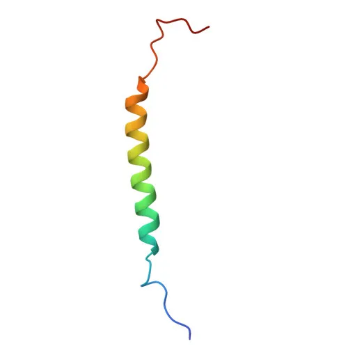
Polyclonal Antibodies
Anti-LAMP2 Polyclonal Antibody (KO Validated) for WB, IHC-P, ELISA - P13473
Item Number : CM0015401
Price varies based on specs and customizations
- Application
- WB, IHC-P, ELISA
- Cross Reactivity
- Human, Mouse
- Protein Weight
- 45kDa

Shipping:
Contact us to get shipping details Enjoy On-time Dispatch Guarantee.
Why Choose Us
Easy ordering process, quality products, and dedicated support for your business success.
Core Product Specifications and Parameters
| Parameter | Value |
|---|---|
| Product Name | [KO Validated] LAMP2 Rabbit pAb |
| Remarks/Alias | DND; LAMPB; CD107b; LAMP-2; LGP-96; LGP110; P2 |
| Species | Human |
| Gene ID | 3920 |
| Immunogen | Recombinant fusion protein containing a sequence corresponding to amino acids 29-375 of human LAMP2 (NP_002285.1). |
| Source | Rabbit |
| Category | Polyclonal Antibodies |
| Application | WB, IHC-P, ELISA |
| Cross Reactivity | Human, Mouse |
| SWISS | P13473 |
| Protein Weight | 45kDa |
| Shipping | Ice bag |
Biological Background: LAMP2 Function and Localization
- LAMP2 (lysosome-associated membrane glycoprotein 2), also designated CD107b, LGP-96, is a highly glycosylated transmembrane protein predominantly localized to lysosomal and endosomal membranes.
- Functions as a key regulator of lysosomal pH by directly inhibiting the proton channel TMEM175, thereby facilitating lysosomal acidification essential for hydrolase activity.
Related references:
PMID:37390818 - Plays a central role in chaperone-mediated autophagy (CMA) by binding substrate proteins (e.g., GAPDH, GPX4) recognized by HSC70 chaperones and targeting them for lysosomal degradation.
Related references:
PMID:11082038
PMID:18644871
PMID:24880125 - Essential for autophagosome-lysosome fusion; LAMP2-deficient cells fail to accumulate syntaxin 17 (STX17) on autophagosomes, blocking autophagic flux.
Related references:
PMID:27628032 - Three major isoforms (LAMP-2A, LAMP-2B, LAMP-2C) are produced by alternative splicing, each with distinct tissue distribution: LAMP-2A is abundant in placenta, lung, and liver; LAMP-2B is broadly expressed; and LAMP-2C is enriched in small intestine, colon, heart, brain, and skeletal muscle.
- Subcellularly, LAMP2 is found on the membrane of lysosomes, late endosomes, the cell surface, and autophagosomes, consistent with its trafficking and roles in autophagy.
- Supports the post-translational processing of certain viral glycoproteins; for instance, it interacts with FURIN to promote cleavage of mumps virus fusion protein F.
Experimental Guidance and Technical Tips
- The immunogen corresponds to amino acids 29–375 of the luminal domain of human LAMP2, a region conserved in mouse, enabling cross-reactivity in both species.
- For western blotting, a band around 45–110 kDa may be observed due to heavy N- and O-linked glycosylation; reducing conditions and deglycosylation enzymes can help confirm specificity.
- In immunohistochemistry (IHC-P), consider using protease-induced antigen retrieval and validate staining in tissues with high LAMP2 expression (e.g., placenta, liver).
- As a polyclonal antibody, it may recognize multiple LAMP2 isoforms; KO validation confirms specificity and is recommended for critical experiments.
- For ELISA applications, the antibody can be employed as both capture and detection reagent after appropriate titration and pairing with a suitable detection system.
CamelBio: Your One-Stop Sourcing Bridge
CamelBio partners with diagnostic manufacturers and research laboratories to provide a comprehensive portfolio of validated IVD raw materials, including polyclonal and monoclonal antibodies, recombinant proteins, and ancillary reagents. This anti-LAMP2 polyclonal antibody supports autophagy and lysosomal research programs and can be sourced alongside complementary detection tools, bulk quantities, and custom antibody development services to accelerate your assay development from concept to clinic.
Product Datasheet
Anti-LAMP2 Polyclonal Antibody (KO Validated) for WB, IHC-P, ELISA - P13473
REQUEST A QUOTE
Our professional team will reply to you within one business day. Please feel free to contact us!