Polyclonal Antibodies
Anti-L-Plastin/LCP1 Polyclonal Antibody for WB, IP, ELISA - P13796
Item Number : CM0030189
Price varies based on specs and customizations
- Application
- WB, IP, ELISA
- Cross Reactivity
- Human, Mouse
- Protein Weight
- 70kDa

Shipping:
Contact us to get shipping details Enjoy On-time Dispatch Guarantee.
Why Choose Us
Easy ordering process, quality products, and dedicated support for your business success.
Core Product Specifications and Parameters
| Parameter | Value |
|---|---|
| Product Name | L-Plastin/LCP1 Rabbit pAb |
| Remarks/Alias | LPL; CP64; PLS2; LC64P; HEL-S-37; L-PLASTIN; L-Plastin/LCP1 |
| Species | Human |
| GeneID | 3936 |
| Immunogen | Recombinant fusion protein containing a sequence corresponding to amino acids 1-270 of human L-Plastin/LCP1 (NP_002289.2). |
| Source | Rabbit |
| Category | Polyclonal Antibodies |
| Application | WB, IP, ELISA |
| Cross Reactivity | Human, Mouse |
| SWISS | P13796 |
| Protein Weight | 70kDa |
| Shipping | Ice bag |
Biological Background: L-Plastin/LCP1 Function and Localization
- L-Plastin/LCP1, also known as Plastin-2 or Lymphocyte cytosolic protein 1 (LCP-1), is an actin-binding protein encoded by the LCP1 gene. Alternative splicing generates multiple isoforms.
- It plays a critical role in actin filament bundling and cytoskeletal organization. Related references: PMID:16636079 PMID:17294403 PMID:28493397
- In T-cells, LCP1 is involved in activation upon costimulation through TCR/CD3 and CD2 or CD28, and modulates the surface expression of IL2RA/CD25 and CD69. Related references: PMID:17294403
- Subcellular localization includes cytoplasm, cytoskeleton, cell junctions, and cell projections such as ruffle membranes.
- Highly expressed in lymphoid organs (spleen, lymph nodes) and immune cells (T-cells, B-cells, monocytes, neutrophils); also detected in intestinal microvilli, hair cell stereocilia, and fibroblast filopodia.
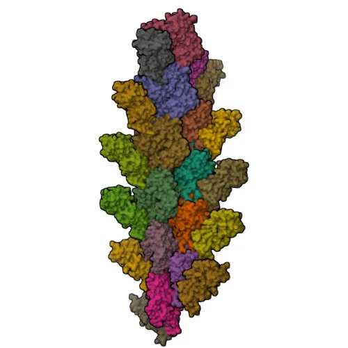
- Post-translational modifications include phosphorylation, acetylation, and calcium binding. The protein is a reference proteome member with a known 3D structure.
- Keywords highlight involvement in chromosomal rearrangements and disease variants, suggesting relevance in pathological conditions.
Experimental Guidance and Technical Tips
- The immunogen corresponds to the N-terminal region (aa 1–270) of human LCP1; the antibody is expected to recognize epitopes within this domain. Validate specificity in your model system.
- For Western blot, human and mouse T-cell or leukocyte lysates are appropriate positive controls given the tissue expression. The predicted band size is ~70 kDa.
- Immunoprecipitation (IP) may be performed using this polyclonal antibody; optimize antibody concentration and incubation conditions for your samples.
- ELISA applications can be explored; consider pairing with a detection antibody if used in sandwich format.
CamelBio: Your One-Stop Sourcing Bridge
CamelBio is your one-stop partner for IVD raw-material sourcing, offering a comprehensive portfolio of antibodies and reagents for diagnostic development. This anti-L-Plastin/LCP1 polyclonal antibody is part of our expanding range for immunology and oncology research. We supply validated antibody pairs, optimized monoclonal and polyclonal antibodies, bulk ancillary reagents, and can assist in sourcing rare-target raw materials to accelerate your assay development from concept to clinic.
REQUEST A QUOTE
Our professional team will reply to you within one business day. Please feel free to contact us!