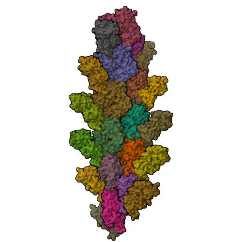
Monoclonal Antibodies
Anti-L-Plastin/LCP1 Monoclonal Antibody for WB, IHC-P, ELISA - P13796
Item Number : CM0011135
Price varies based on specs and customizations
- Application
- WB, IHC-P, ELISA
- Cross Reactivity
- Human
- Protein Weight
- 70kDa

Shipping:
Contact us to get shipping details Enjoy On-time Dispatch Guarantee.
Why Choose Us
Easy ordering process, quality products, and dedicated support for your business success.
Core Product Specifications and Parameters
| Parameter | Value |
|---|---|
| Product Name | L-Plastin/LCP1 Rabbit mAb |
| Remarks/Alias | LPL; CP64; PLS2; LC64P; HEL-S-37; L-PLASTIN |
| Species | Human |
| Gene ID (Human) | 3936 |
| Gene ID | 3936 |
| Immunogen | A synthetic peptide corresponding to a sequence within amino acids 528-627 of human L-Plastin/LCP1 (P13796). |
| Source | Rabbit |
| Category | Monoclonal Antibodies |
| Application | WB, IHC-P, ELISA |
| Cross Reactivity | Human |
| SWISS | P13796 |
| Protein Weight | 70kDa |
| Shipping | Ice bag |
Biological Background: L-Plastin/LCP1 Function and Localization
- L-Plastin/LCP1, also known as Plastin-2 or Lymphocyte cytosolic protein 1 (LCP-1), is an actin-binding protein encoded by the LCP1 gene (alternative splicing yields additional isoforms).
- It plays a critical role in T-cell activation by modulating the cell surface expression of IL2RA/CD25 and CD69 upon costimulation via TCR/CD3 and CD2 or CD28. Related references: PMID:17294403
- L-Plastin/LCP1 binds to actin filaments, facilitating cytoskeletal reorganization essential for immune cell function and cellular motility. Related references: PMID:16636079 PMID:28493397
- The protein localizes primarily to the cytoplasm and cytoskeleton, and is enriched at cell junctions, cell projections, and ruffle membranes, consistent with its role in cell shape and migration.
- It is highly expressed in immune tissues (spleen, lymph nodes) and in hematopoietic cells including T-lymphocytes, neutrophils, monocytes, B-lymphocytes, and myeloid cells, as well as in intestinal microvilli, hair cell stereocilia, and fibroblast filopodia.
- Post-translational modifications include phosphorylation, acetylation, and calcium binding, which regulate its actin-bundling activity and subcellular dynamics.
- Reflecting its multifaceted roles, L-Plastin/LCP1 is associated with cellular processes such as chromosomal rearrangement, disease variants, and is identified through extensive proteomic studies.
Experimental Guidance and Technical Tips
- The immunogen is a synthetic peptide corresponding to the C-terminal region (aa 528–627) of human L-Plastin/LCP1, suggesting the antibody likely recognizes an epitope in this region. For Western blot, a band around 70 kDa is expected; consider using a positive control such as human lymphoid or myeloid cell lysates.
- For immunohistochemistry (paraffin), antigen retrieval may be optimized for detection in tissues with high L-Plastin expression, such as spleen or lymph node sections.
- In ELISA applications, the rabbit monoclonal format provides high specificity; validate in your specific sample matrix to confirm linearity and sensitivity.
- Because L-Plastin/LCP1 is involved in T-cell activation, consider using this antibody to study signaling pathways in stimulated vs. unstimulated immune cells.
CamelBio: Your One-Stop Sourcing Bridge
CamelBio provides diagnostic manufacturers, laboratories, and research institutes with one-stop access to IVD raw materials, technical services, and consulting—covering every stage from concept to clinic. Our portfolio includes validated antibody pairs, optimized monoclonal/polyclonal antibodies, bulk ancillary reagents, and rare target raw-material sourcing tailored to your development needs. For research into immune function and cytoskeletal disorders, this anti-L-Plastin/LCP1 monoclonal antibody is one of many high-quality reagents we offer to accelerate your assay development.
Product Datasheet
Anti-L-Plastin/LCP1 Monoclonal Antibody for WB, IHC-P, ELISA - P13796
REQUEST A QUOTE
Our professional team will reply to you within one business day. Please feel free to contact us!