Polyclonal Antibodies
Anti-KLRK1 Rabbit Polyclonal Antibody for IF/ICC and ELISA - P26718
Item Number : CM0018331
Price varies based on specs and customizations
- Application
- IF/ICC, ELISA
- Cross Reactivity
- Human
- Protein Weight
- 25kDa

Shipping:
Contact us to get shipping details Enjoy On-time Dispatch Guarantee.
Why Choose Us
Easy ordering process, quality products, and dedicated support for your business success.
Core Product Specifications and Parameters
| Parameter | Value |
|---|---|
| Product Name | KLRK1 Rabbit pAb |
| Remarks/Alias | KLR; CD314; NKG2D; NKG2-D; D12S2489E; KLRK1 |
| Species | Human |
| Gene ID (Human) | 22914 |
| Gene ID | 22914 |
| Immunogen | Synthetic peptide corresponding to a sequence within amino acids 50-150 of human KLRK1 (NP_031386.2) |
| Source | Rabbit |
| Category | Polyclonal Antibodies |
| Application | IF/ICC, ELISA |
| Cross Reactivity | Human |
| SWISS | P26718 |
| Protein Weight | 25kDa |
| Shipping | Ice bag |
Biological Background: KLRK1/NKG2D Function and Localization
- KLRK1 (also known as NKG2D, CD314) encodes the NKG2-D type II integral membrane protein, a killer cell lectin-like receptor subfamily K member 1. It functions as an activating and costimulatory receptor central to innate and adaptive immunosurveillance.
- Upon binding to stress-inducible MHC class I-related ligands (e.g., MICA/B, ULBP1-6) on tumor or virus-infected cells, NKG2D delivers stimulatory signals that trigger calcium influx, culminating in TNF-alpha expression and perforin-mediated cytotoxicity.
- In natural killer (NK) cells, NKG2D provides both primary activation and costimulatory signals, while in CD8+ T cells it acts as a costimulatory receptor amplifying TCR-mediated adaptive responses.
- The receptor is expressed on essentially all CD56+CD3- NK cells from peripheral blood, as well as on CD8+ alpha-beta and gamma-delta T cells and interferon-producing killer dendritic cells (IKDCs).
- Subcellularly, NKG2D localizes to the cell membrane as a transmembrane protein with a signal-anchor topology. It is subject to glycosylation and contains a disulfide bond within its extracellular ligand-binding domain.
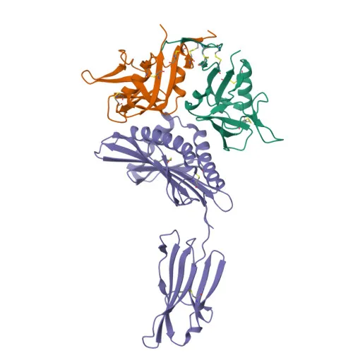
- Structural studies (3D-structure) have elucidated the binding interface with ligands, and the protein is a reference proteome member with alternative splicing generating additional isoforms.
Experimental Guidance and Technical Tips
- The immunogen was a synthetic peptide spanning amino acids 50–150 of human KLRK1 (NP_031386.2). For proper recognition, ensure the antibody is applied to samples where the target sequence is preserved and the epitope is accessible (e.g., non-denaturing conditions for IF/ICC).
- Validate the antibody in your specific cellular context using positive controls such as IL-2-activated NK cells or CD8+ T-cell lines known to express KLRK1. For ELISA, consider pairing with a capture antibody if developing a sandwich immunoassay.
- Although not tested here, KLRK1 is a transmembrane protein with an extracellular ligand-binding domain; consider cell surface staining protocols that avoid permeabilization for intact membrane detection in flow cytometry experiments.
CamelBio: Your One-Stop Sourcing Bridge
CamelBio provides diagnostic manufacturers and research labs with streamlined access to high-quality IVD raw materials, from validated antibody pairs and monoclonal/polyclonal antibodies to bulk ancillary reagents. For researchers focused on immuno-oncology and NK cell biology, we offer customized sourcing for rare targets like KLRK1—whether you need matched detection pairs for ELISA development or consistent production batches for assay scale-up. Partner with CamelBio to accelerate your project from concept to clinic with reliable, well-documented raw materials.
REQUEST A QUOTE
Our professional team will reply to you within one business day. Please feel free to contact us!