Polyclonal Antibodies
Anti-Klotho Polyclonal Antibody for WB and ELISA - Q9UEF7
Item Number : CM0018685
Price varies based on specs and customizations
- Application
- WB, ELISA
- Cross Reactivity
- Human, Mouse, Rat
- Protein Weight
- 116kDa

Shipping:
Contact us to get shipping details Enjoy On-time Dispatch Guarantee.
Why Choose Us
Easy ordering process, quality products, and dedicated support for your business success.
Core Product Specifications and Parameters
| Parameter | Value |
|---|---|
| Product Name | KL Rabbit pAb |
| Remarks/Alias | KLA; HFTC3; KL |
| Species | Human |
| GeneID (Human) | 9365 |
| GeneID | 9365 |
| Immunogen | Recombinant fusion protein containing a sequence corresponding to amino acids 1-200 of human KL (NP_004786.2). |
| Source | Rabbit |
| Category | Polyclonal Antibodies |
| Application | WB, ELISA |
| Cross Reactivity | Human, Mouse, Rat |
| SWISS | Q9UEF7 |
| Protein Weight | 116kDa |
| Shipping | Ice bag |
Biological Background: Klotho Function and Localization
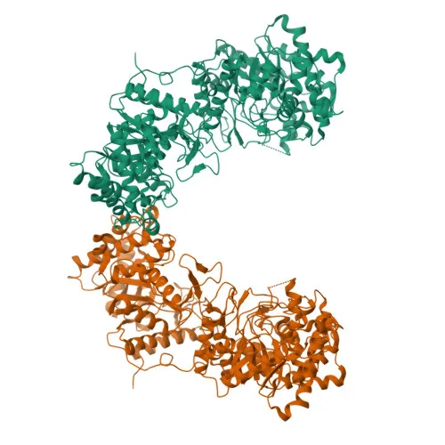
- Klotho (KL) is a single-pass type I membrane protein, encoded by the KL gene in humans, and also exists as a secreted peptide generated by proteolytic cleavage.
- It exhibits beta-glucuronidase activity, capable of hydrolyzing steroid beta-D-glucuronides such as estradiol-3-beta-D-glucuronide, estrone-3-beta-D-glucuronide, and estriol-3-beta-D-glucuronide.
- Klotho acts as an obligate co-receptor for FGF23, essential for fibroblast growth factor 23 (FGF23) binding to FGFR1 and mediating FGF23-regulated renal vitamin D metabolism and phosphate reabsorption.
- The shed soluble Klotho ectodomain functions as a circulating anti-aging hormone that may extend lifespan by inhibiting insulin/IGF1 signaling.
- Subcellular localization includes the apical cell membrane (membrane-bound isoform) and secretion (soluble isoform), allowing autocrine, paracrine, and endocrine functions.
- Tissue expression is highest in kidney, placenta, small intestine, and prostate; soluble Klotho is present in serum and cerebrospinal fluid and is downregulated in certain cancers and chronic kidney disease.
- Post-translational modifications include glycosylation, and the protein is classified under glycosidase, hormone, and hydrolase families.
Experimental Guidance and Technical Tips
- The immunogen used spans the N-terminal region (aa 1–200) of human Klotho, which is common to both membrane and secreted isoforms; consider this when designing detection assays.
- For Western blot (WB), Klotho migrates around 116 kDa; include positive controls from kidney, placenta, or prostate lysates (human, mouse, or rat) as these tissues express the protein.
- When performing ELISA, validate antibody specificity using recombinant Klotho protein or biological fluids where soluble Klotho is abundant, such as serum or cerebrospinal fluid.
- Cross-reactivity with mouse and rat Klotho is predicted; however, sequence alignment and pilot testing are recommended to confirm similar reactivity in these species.
- Optimize antibody dilution and detection conditions for each application; typical starting dilutions should be empirically determined.
CamelBio: Your One-Stop Sourcing Bridge
CamelBio provides diagnostic manufacturers and research institutions with one-stop access to IVD raw materials, technical services, and consulting for every stage from concept to clinic. The Klotho polyclonal antibody supports research into aging, renal physiology, and endocrine signaling, with our supply capabilities encompassing validated antibody pairs, optimized monoclonal/polyclonal reagents, bulk ancillary materials, and rare-target raw-material sourcing. We offer efficient procurement and tailored support to streamline your assay development and manufacturing workflows.
REQUEST A QUOTE
Our professional team will reply to you within one business day. Please feel free to contact us!