Polyclonal Antibodies
Anti-KLK6 Polyclonal Antibody for WB, ELISA - Q92876
Item Number : CM0016327
Price varies based on specs and customizations
- Application
- WB, ELISA
- Cross Reactivity
- Human
- Protein Weight
- 26kDa

Shipping:
Contact us to get shipping details Enjoy On-time Dispatch Guarantee.
Why Choose Us
Easy ordering process, quality products, and dedicated support for your business success.
Core Product Specifications and Parameters
| Parameter | Value |
|---|---|
| Product Name | KLK6 Rabbit pAb |
| Remarks/Alias | hK6; Bssp; Klk7; SP59; PRSS9; PRSS18; KLK6 |
| Species | Human |
| Gene ID | 5653 |
| Immunogen | Synthetic peptide corresponding to a sequence within amino acids 100 to the C-terminus of human KLK6 (NP_002765.1) |
| Source | Rabbit |
| Category | Polyclonal Antibodies |
| Application | WB, ELISA |
| Cross Reactivity | Human |
| SWISS | Q92876 |
| Protein Weight | 26kDa |
| Shipping | Ice bag |
Biological Background: KLK6 Function and Localization
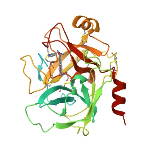
- Kallikrein-6 (KLK6), also known as neurosin, protease M, SP59, or zyme, is a secreted serine protease encoded by the KLK6 gene (alternative names PRSS18/PRSS9), belonging to the tissue kallikrein family.
- KLK6 preferentially cleaves substrates with Arg at the P1 position and Ser/Pro at P2, exhibiting activity against amyloid precursor protein, myelin basic protein, gelatin, casein, and extracellular matrix proteins (fibronectin, laminin, vitronectin, collagen).
- The protease degrades alpha-synuclein and inhibits its polymerization, suggesting a protective role in synucleinopathies such as Parkinson disease; its dysregulation may contribute to pathogenesis.
- In the nervous system, KLK6 is implicated in regulating axon outgrowth after spinal cord injury, and it localizes to gray matter neurons where it colocalizes with pathological inclusions like Lewy bodies and glial cytoplasmic inclusions.
- Overexpressed in primary breast tumors but not metastatic tumors, KLK6 promotes invasion and metastasis, as tumor cells treated with a neutralizing antibody show reduced migration.
- Subcellular localization includes secretion, nucleus/nucleolus, cytoplasm, mitochondria, and microsomes.
- Post-translational modifications encompass glycosylation, disulfide bond formation, autocatalytic cleavage, and processing from a zymogen to an active serine protease.
- Tissue expression is highest in fluids (milk, cerebrospinal fluid) and glandular tissues (salivary glands, lung, colon, breast, kidney) and detected in brain gray matter; it is not found in several other tissues including skin, spleen, liver, and pancreas.
Experimental Guidance and Technical Tips
- The immunogen targets the C-terminal region (aa 100–C-terminus) of human KLK6, which may recognize both zymogen and active forms. Confirm specificity with appropriate controls (recombinant protein, knockdown/knockout lysates).
- For Western blot, the predicted molecular weight is ~26 kDa; glycosylation may cause a higher apparent band. Use positive controls (human milk or cerebrospinal fluid) and consider reducing conditions to break disulfide bonds.
- For ELISA, optimize coating antigen and antibody dilution. The antibody can be used as capture or detection reagent in sandwich ELISA; assess cross-reactivity with other kallikreins.
- KLK6 is secreted; test cell culture supernatants or biological fluids (CSF, milk) alongside lysates. Minimize freeze-thaw cycles to preserve protein integrity.
CamelBio: Your One-Stop Sourcing Bridge
CamelBio provides diagnostic manufacturers, labs, and research institutes with one-stop access to IVD raw materials, technical services, and consulting from concept to clinic. For KLK6-focused projects, we offer validated antibody pairs, optimized monoclonal/polyclonal antibodies, bulk ancillary reagents, and rare target raw-material sourcing. Our team supports neurodegenerative and oncology biomarker development with comprehensive supply-chain solutions.
REQUEST A QUOTE
Our professional team will reply to you within one business day. Please feel free to contact us!