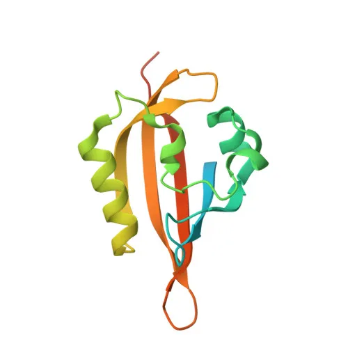
Polyclonal Antibodies
Anti-KCNH1 Polyclonal Antibody for WB, ELISA - O95259
Item Number : CM0028629
Price varies based on specs and customizations
- Application
- WB, ELISA
- Cross Reactivity
- Human, Mouse, Rat
- Protein Weight
- 111kDa

Shipping:
Contact us to get shipping details Enjoy On-time Dispatch Guarantee.
Why Choose Us
Easy ordering process, quality products, and dedicated support for your business success.
Core Product Specifications and Parameters
| Specification | Detail |
|---|---|
| Product Name | KCNH1 Rabbit pAb |
| Remarks/Alias | EAG; EAG1; ZLS1; hEAG; TMBTS; h-eag; hEAG1; Kv10.1; KCNH1 |
| Species | Human |
| GeneID (Human) | 3756 |
| GeneID | 3756 |
| Immunogen | Recombinant fusion protein containing a sequence corresponding to amino acids 813-962 of human KCNH1 (NP_002229.1) |
| Source | Rabbit |
| Category | Polyclonal Antibodies |
| Application | WB, ELISA |
| Cross Reactivity | Human, Mouse, Rat |
| SWISS | O95259 |
| Protein Weight | 111kDa |
| Shipping | Ice bag |
Biological Background: KCNH1 Function and Localization
- KCNH1 encodes the pore-forming alpha subunit of a voltage-gated delayed rectifier potassium channel that mediates outward-rectifying potassium currents which do not inactivate upon depolarization (PubMed:10880439, PubMed:22732247).
Related references: PMID:10880439 PMID:22732247 - Channel activation kinetics are dependent on prepulse potential and external divalent cation concentration, exhibiting a biphasic time course with fast and slowly activating components (PubMed:11943152).
Related references: PMID:11943152 - Channel properties are modulated by subunit assembly (PubMed:11943152).
Related references: PMID:11943152 - KCNH1 mediates IK(NI) current in myoblasts (PubMed:9738473).
Related references: PMID:9738473 - Involved in the regulation of cell proliferation and differentiation, particularly adipogenic and osteogenic differentiation in bone marrow-derived mesenchymal stem cells (MSCs) (PubMed:23881642).
Related references: PMID:23881642 - KCNH1 localizes to the cell membrane, nuclear inner membrane, dendrites, axons, presynaptic and postsynaptic membranes, perikaryon, and early endosome membrane.
- Highly expressed in brain and in myoblasts at the onset of fusion, with detection in certain cancer cell lines (e.g., HeLa, SH-SY5Y, MCF-7) but not in normal epithelial cells.
Experimental Guidance and Technical Tips
- The immunogen comprises amino acids 813-962, a C-terminal region of human KCNH1. This polyclonal antibody may recognize the full-length protein (~111 kDa) in Western blot assays; consider using reducing SDS-PAGE conditions.
- For ELISA applications, validate the antibody against the recombinant immunogen or full-length protein under your experimental conditions.
- Given the reported cross-reactivity with human, mouse, and rat, this antibody can be evaluated in rodent models. Test in relevant tissues such as brain or myoblast cell lines.
- KCNH1 exhibits complex subcellular localization; fixation and permeabilization protocols should be optimized if using this antibody for immunostaining.
- Include appropriate positive and negative controls (e.g., brain lysate, myoblast lysate, KCNH1 knockout samples) to confirm specificity.
CamelBio: Your One-Stop Sourcing Bridge
CamelBio offers a comprehensive sourcing solution for IVD raw materials, including high-quality antibodies like this anti-KCNH1 polyclonal antibody. We support diagnostic manufacturers and researchers with validated antibody pairs, optimized monoclonal/polyclonal antibodies, bulk ancillary reagents, and customized raw material sourcing for rare targets. Leverage our expertise to accelerate your assay development from concept to clinic.
REQUEST A QUOTE
Our professional team will reply to you within one business day. Please feel free to contact us!