Polyclonal Antibodies
Anti-KANK2 Rabbit Polyclonal Antibody for WB, IHC-P, ELISA - Q63ZY3
Item Number : CM0021372
Price varies based on specs and customizations
- Application
- WB, IHC-P, ELISA
- Cross Reactivity
- Human, Mouse, Rat
- Protein Weight
- 91kDa

Shipping:
Contact us to get shipping details Enjoy On-time Dispatch Guarantee.
Why Choose Us
Easy ordering process, quality products, and dedicated support for your business success.
Core Product Specifications and Parameters
| Parameter | Value |
|---|---|
| Product Name | KANK2 Rabbit pAb |
| Remarks/Alias | SIP; MXRA3; PPKWH; NPHS16; ANKRD25; KANK2 |
| Species | Human |
| Gene ID (Human) | 25959 |
| Gene ID | 25959 |
| Immunogen | Recombinant fusion protein containing a sequence corresponding to amino acids 1-300 of human KANK2 (NP_001129663.1). |
| Source | Rabbit |
| Category | Polyclonal Antibodies |
| Application | WB, IHC-P, ELISA |
| Cross Reactivity | Human, Mouse, Rat |
| SWISS | Q63ZY3 |
| Protein Weight | 91kDa |
| Shipping | Ice bag |
Biological Background: KANK2 Function and Localization
- KANK2 (KN motif and ankyrin repeat domain-containing protein 2), also known as ANKRD25, MXRA3, or SIP, is a scaffold protein with multiple roles in transcription regulation, apoptosis, and cytoskeletal dynamics.
- Acts as a transcriptional regulator by sequestering nuclear receptor coactivators NCOA1, NCOA2, and NCOA3 in the cytoplasm, thereby modulating gene expression. Related references: PMID:17476305
- Plays a key role in caspase-independent apoptosis by retaining the proapoptotic factor AIFM1 in mitochondria; upon proteasomal degradation of KANK2, AIFM1 translocates to the nucleus to induce cell death. Related references: PMID:22371500
- Negatively regulates vitamin D receptor (VDR) signaling, influencing calcium homeostasis and cell differentiation pathways. Related references: PMID:24671081
- Promotes actin stress fiber formation and cell adhesion/migration through interaction with ARHGDIA and modulation of Rho GTPase signaling; involved in podocyte migration during kidney development. Related references: PMID:17996375 | PMID:25961457
- Localizes to both the cytoplasm and mitochondria, consistent with its dual roles in cytoskeletal organization and apoptosis.
- Strongly expressed in cervix, colon, heart, kidney, and lung; at the protein level, detected in kidney glomerular podocytes and mesangial cells.
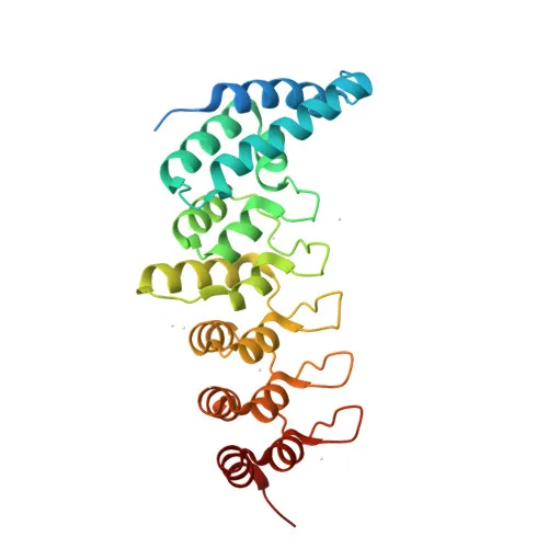
- Contains multiple ANK repeats and a coiled-coil domain; undergoes alternative splicing, phosphorylation, and methylation, and is associated with diseases such as palmoplantar keratoderma and nephrotic syndrome type 16 (NPHS16).
Experimental Guidance and Technical Tips
- The immunogen encompasses amino acids 1–300 of human KANK2, covering an N-terminal region that is conserved across species; consider this when assessing cross-reactivity.
- For western blotting, expect a band near 91 kDa; validate with appropriate positive and negative controls (e.g., cell lines expressing KANK2).
- In IHC-P, optimize antigen retrieval and titre using human kidney or colon sections known to exhibit strong KANK2 expression.
- For ELISA, the recombinant immunogen may serve as a calibration standard; test a concentration range to establish assay linearity.
- As a polyclonal antibody, lot-to-lot consistency should be monitored; always include a control standard in each experiment.
CamelBio: Your One-Stop Sourcing Bridge
CamelBio serves as a comprehensive sourcing partner for IVD raw materials, offering validated antibody pairs, optimized monoclonal and polyclonal antibodies, bulk ancillary reagents, and tailored solutions for rare targets. Our KANK2 antibody supports research into transcriptional regulation, apoptosis, and kidney development, and we can assist with custom manufacturing, technical consultation, and seamless integration into diagnostic workflows.
REQUEST A QUOTE
Our professional team will reply to you within one business day. Please feel free to contact us!