Monoclonal Antibodies
Anti-ITPR3 Rabbit Monoclonal Antibody for WB, ELISA - Q14573
Item Number : CM0004035
Price varies based on specs and customizations
- Application
- WB, ELISA
- Cross Reactivity
- Human
- Protein Weight
- 304kDa

Shipping:
Contact us to get shipping details Enjoy On-time Dispatch Guarantee.
Why Choose Us
Easy ordering process, quality products, and dedicated support for your business success.
Core Product Specifications and Parameters
| Parameter | Value |
|---|---|
| Product Name | ITPR3 Rabbit mAb |
| Remarks/Alias | IP3R; CMT1J; IP3R3; ITPR3 |
| Species | Human |
| GeneID (Human) | 3710 |
| GeneID | 3710 |
| Immunogen | Recombinant protein containing a sequence corresponding to amino acids 1630-1870 of human ITPR3 (NP_002215.2) |
| Source | Rabbit |
| Category | Monoclonal Antibodies |
| Application | WB, ELISA |
| Cross Reactivity | Human |
| SWISS | Q14573 |
| Protein Weight | 304kDa |
| Shipping | Ice bag |
Biological Background: ITPR3 Function and Localization
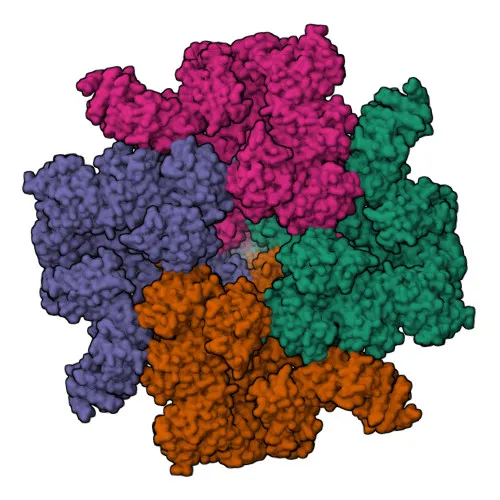
- ITPR3 is an inositol 1,4,5-trisphosphate-gated calcium channel that releases calcium from the endoplasmic reticulum into the cytosol upon ligand binding, playing a key role in intracellular calcium homeostasis. Related references: PMID:32949214, PMID:37898605, PMID:8081734
- The channel undergoes a multi-step activation process: IP3 binding induces a resting-to-preactivated transition, increasing calcium affinity, which then promotes opening via high-energy intermediate states, with calcium binding at distinct sites regulating both activation and inhibition. Related references: PMID:30013099, PMID:35301323, PMID:37898605
- ITPR3 is also known as IP3 receptor isoform 3 (IP3R3) or Type 3 InsP3 receptor, encoded by the ITPR3 gene.
- Subcellularly, it localizes primarily to the endoplasmic reticulum membrane and to cytoplasmic secretory vesicle membranes.
- Tissue expression is observed in intestinal crypt and villus epithelial cells.
- The protein contains transmembrane helices and repeat domains, and is classified as a ligand-gated ion channel involved in calcium transport; it is subject to phosphorylation and contains structural disulfide bonds.
- Genetic variants in ITPR3 are associated with Charcot-Marie-Tooth disease and ectodermal dysplasia.
- The full-length protein has a molecular weight of approximately 304 kDa.
Experimental Guidance and Technical Tips
- The immunogen corresponds to amino acids 1630–1870 in the C‑terminal region of human ITPR3. When setting up WB, anticipate a band near 304 kDa and consider using a low‑percentage gel and extended transfer times to ensure efficient blotting of this large protein.
- For ELISA applications, validating the antibody against the recombinant immunogen or full‑length protein is recommended; a sandwich ELISA format may require pairing with a detection antibody recognizing a separate epitope.
- Because cross‑reactivity is reported only for human, verify reactivity in your specific sample type (e.g., cell lines or tissue lysates) and consider using appropriate positive and negative controls.
- To confirm specificity, a knockdown or knockout control in the relevant cell system is advisable, as the epitope may share homology with other IP3 receptor isoforms.
CamelBio: Your One-Stop Sourcing Bridge
CamelBio provides diagnostic manufacturers and researchers with streamlined access to a comprehensive range of IVD raw materials, including this ITPR3 monoclonal antibody for calcium signaling research. Our portfolio encompasses validated antibody pairs, optimized monoclonal and polyclonal antibodies, and bulk ancillary reagents. For any rare-target raw-material needs, our expert sourcing network helps accelerate your assay development from concept to clinic.
REQUEST A QUOTE
Our professional team will reply to you within one business day. Please feel free to contact us!